New evidence links brain geometry to cognitive decline
A recent multi-institution study indicates that subtle changes in the three-dimensional shape of the brain may signal future cognitive decline and dementia risk. Researchers analyzed thousands of MRI scans to map how brain geometry evolves with age and how those shifts correlate with declines in memory, reasoning and other cognitive functions.
The research, led by teams at the University of California, Irvine (UC Irvine) and the University of La Laguna in Spain, expands the focus from simple tissue loss to the brain’s overall morphology — the spatial arrangement and curvature of distinct regions. Instead of asking only how much volume the brain loses, the investigators looked at how the brain’s surface and internal structure deform over time and whether those deformations align with poorer cognitive test performance.
Study design and methods
The authors analyzed 2,603 MRI brain scans from adults aged 30 to 97, tracking shape and structural changes across specific regions and comparing those patterns with standardized cognitive assessments. Advanced image-processing and statistical mapping allowed the team to identify areas that consistently expanded or contracted with age, and to test whether those patterns were stronger in people showing measurable cognitive impairment.
Key methodological points
- Large, cross-sectional and longitudinal MRI dataset covering mid-adulthood to very old age.
- Regional shape modeling rather than global volume measures alone.
- Cognitive scores used to stratify participants by performance on memory, reasoning and other domains.
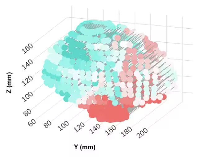
The study produced regional maps of age-related expansions and contractions in brain geometry and linked these to cognitive test scores. The authors presented these maps and analytic results in their paper. (Escalante et al., Nat. Commun., 2025)
Main findings and implications
The spatial changes were not uniform: posterior (rear) regions of the brain tended to show more age-related shrinkage, and those reductions were associated with lower reasoning scores. In some individuals, the pattern of uneven expansion and compression across brain regions was more pronounced and correlated with cognitive impairment. These findings suggest that the distribution of structural change — not just the total volume lost — matters for brain health.
A particularly important implication concerns the entorhinal cortex, a compact region that acts as a key hub for memory networks and is one of the first areas affected by Alzheimer’s pathology. The researchers propose that age-related shifts in adjacent brain geometry could mechanically stress the entorhinal cortex against nearby rigid structures, potentially making it more vulnerable to the accumulation of toxic proteins linked to Alzheimer's disease.
Neuroscientist Niels Janssen (University of La Laguna) summarized the shift in perspective: rather than focusing solely on how much tissue disappears with age, it is important to map how the brain’s shape changes in systematic ways and how those shifts relate to cognition. Michael Yassa (UC Irvine) added that such geometric considerations may help explain why the entorhinal cortex often becomes 'ground zero' for Alzheimer’s pathology and could offer new routes for early detection and intervention.
Future directions and research needs
The study is an early but important step. Researchers emphasize the need for larger longitudinal datasets, higher-resolution imaging, and integration with molecular markers (for example, PET scans measuring amyloid and tau) to determine causality: do shape changes precede protein accumulation, or are they a consequence of it? Better biomechanical models and population-diverse datasets will help clarify why some regions expand with age while others contract, and why some people show more pronounced geometric shifts than others.
The clinical prospects include using automated MRI-based shape metrics to flag individuals at elevated dementia risk earlier than conventional volumetric screening. If validated, shape-based biomarkers could complement cognitive testing and biochemical markers to refine diagnosis and timing of interventions.
Expert Insight
Dr. Laura Mendes, a cognitive neuroscientist (fictional), commented: "This research reframes aging as not only a problem of tissue loss but of architectural change. Brain regions are embedded in a physical space; when that space remodels, vulnerable hubs can be strained. Combining geometry with molecular imaging could be a game-changer for early diagnosis."
Conclusion
This study highlights that the 3D geometry of the brain — its shape and regional deformation patterns — carries meaningful information about cognitive aging and dementia risk. Tracking those shape changes alongside established biomarkers may open new paths for early detection and targeted care strategies.

“My work centers on sustainability, energy, and environmental science — examining how innovation can lead to a greener future.”

Discussion
Leave a Comment