Something small and unexpected has turned a familiar chapter of Earth’s deep past sideways. A battered skull, misread for years, and a pristine specimen from Yunnan are forcing paleontologists to rethink how the fish that eventually led to land-dwelling vertebrates first diversified.
New reconstructions, made possible by modern CT scanning and computed tomography, have pulled anatomical detail out of fossils once written off as too damaged to study. The results lace together stories emerging from two continents: the Devonian reefs of northern Australia and marine deposits in South China. Together they reveal a burst of innovation among early lungfish — the lobe-finned fishes that sit closest to the branch of vertebrates that later walked onto land.
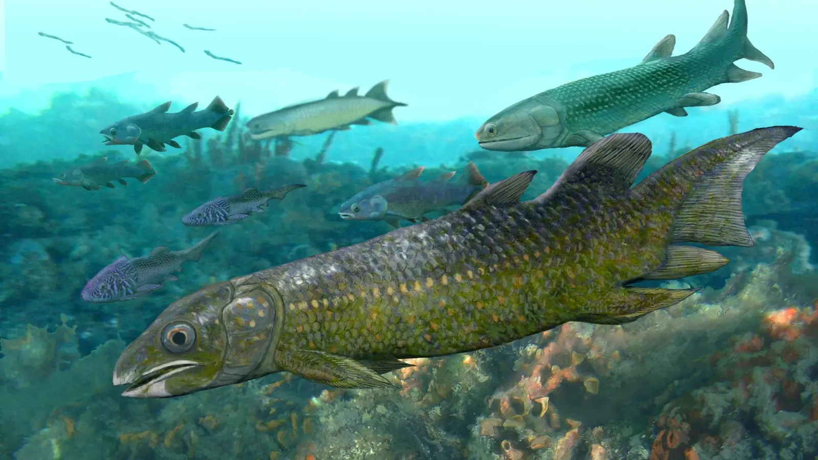
Revealing a misread skull
In the Kimberley region of Western Australia lies the Gogo Formation, a reef complex often called an ancient Great Barrier Reef. For decades it has been a treasure trove for Devonian fossils, yielding exquisitely preserved specimens that capture soft anatomy rarely fossilized. Yet one peculiar fossil from Gogo has haunted researchers: incomplete, distorted, and so odd that the team first describing it in 2010 wondered whether it represented an entirely new kind of fish.
Using high-resolution CT imaging and support from facilities including ANSTO, Flinders University paleontologists re-scanned the specimen and built detailed digital models of its skull and brain cavity. The picture changed. What earlier workers had interpreted from surface impressions was, quite literally, upside down and backward in places. The internal anatomy — especially the inner ear region and the cranial cavity — aligns more closely with known Gogo lungfish than previously thought, while still carrying unusual traits that hint at hidden diversity within the reef community.

3D print of Chirodipterus australis skull, a lungfish from the Gogo Formation, which shares close similarities to Cainocara enigma.
Dr. Alice Clement, who led the reanalysis, says the study doesn’t simply correct an old interpretation; it expands the catalogue of anatomical variation preserved at Gogo. What looked like damage or deformation now provides a new data point on how cranial structures evolved among Devonian lungfish — a group instrumental in the story of tetrapod origins.
A 410-million-year-old snapshot from China
While Australian fossils clarified one mystery, a separate find in South China added another piece to the puzzle. A nearly complete skull described as Paleolophus yunnanensis (sometimes written Paleolopus in popular summaries) dates to roughly 410 million years ago. This specimen represents a transitional form: not the earliest lungfish, but a specimen from the interval when the lineage rapidly developed the bite and jaw mechanics later seen across the Devonian.
Paleolophus reveals a mosaic of traits. Some features echo the primitive Diabolepis fossils known from southern China; others align with mid-Devonian forms described from Wyoming and more derived species recorded in Australia. The result is a clearer view of how feeding adaptations — crushing tooth plates, changes to jaw articulation, and skull reinforcement — emerged and diversified in a relatively short geological window.
Dr. Brian Choo, involved in the Yunnan work, describes the fossil as an unprecedented look at a group in the act of diversification. The Chinese specimen highlights geographical links across ancient Gondwana and Laurussia, suggesting that anatomical experiments among lungfish were widespread, not isolated curiosities.
All of this matters because lungfish are close cousins of the tetrapods. Understanding their cranial and sensory anatomy helps paleontologists infer the suite of changes that made the water-to-land transition possible: shifts in respiration, bite mechanics, and balance systems required for life in shallower, more complex habitats.
Technologies changing fossil narratives
Modern imaging reshapes more than bones. CT scanning can reveal tiny canals in the inner ear that inform how these animals sensed motion and position. It can resolve brain cavity shapes that hint at neural specializations and reproduce delicate laminae of bone that were once lost to erosion. When researchers combine scans from living lungfish with fossilized crania, they can test evolutionary scenarios with a precision previously impossible.
Coauthor Hannah Thiele emphasized that the work is collaborative across museums, imaging centers, and international institutions. It’s a reminder: the story of life’s major chapters is often rewritten not by new field sites, but by fresh looks at old specimens.

Dr. Alice Clement, left, and palaeontology student Hannah Thiele at Flinders University, Australia.
Expert Insight
"Fossils are not static evidence; they are hypotheses waiting to be re-examined," says Dr. Maria López, an evolutionary paleobiologist unaffiliated with the studies. "When imaging reveals internal structure we couldn’t see from the surface, entire phylogenetic placements can shift. That ripple affects how we model the origins of limbs, lungs, and the suite of traits linking fish to tetrapods."
These studies do not close the book on Devonian lungfish. They open new pages. With each scan and each reanalysis, paleontologists get closer to reconstructing not just who these animals were, but how swiftly and repeatedly evolutionary experiments unfolded during a critical interval in Earth’s history. The next seemingly inscrutable fossil may be the one that finally fills another gap in the bridge from water to land.

“The cosmos has always fascinated me. I write about space missions, astronomy, and the technologies pushing humanity beyond Earth.”

Discussion
Leave a Comment