Imagine a miniature heart that not only beats on its own but also tells you, in fine print, what each cell is doing. That’s the promise of the latest heart-on-a-chip (HOC) platform developed by a Canadian research team: a three-dimensional engineered cardiac tissue that pairs bulk force sensing with tiny, tissue-embedded microsensors to watch muscle cells at work.
For decades, cardiology has been limited by the problem that matters most: you can’t safely test how an actual human heart will react to a new drug or a disease without risk. Animal models and simplified cell cultures help, but they miss the nuanced mechanics of contracting heart muscle. This new device fills that gap by combining two complementary measurement systems: macro-scale pillars that bend with each beat, and hydrogel-based microdroplets that report local stresses where cells generate force.
How the dual-sensing heart is built
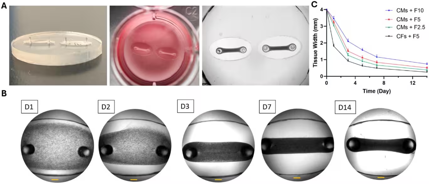
Take stem cells or animal-derived cardiac cells, suspend them in a protein-rich gel, and let them organize into dense muscle tissue. That is the starting point. The researchers used rat cardiomyocytes and connective cells embedded in a supportive extracellular-matrix-like gel, then seeded the mixture onto a silicon chip. The tissue self-assembles into a beating strip suspended between two flexible elastic pillars. Each contraction deflects the pillars; the amount and timing of that deflection reveal whole-tissue contractile strength and rhythm.

But the real novelty is the micro-sensing layer. Tiny hydrogel droplets, about 50 micrometers across, are dispersed inside the tissue. They deform under the very small, local forces produced by individual cardiomyocytes and their neighbors. By tracking droplet shape and motion, the team can infer cellular-level mechanical stresses that are invisible to conventional HOC platforms.
Sensors that bridge scales
The pillar system answers the question: how hard does the engineered heart muscle beat? The microdroplets answer a subtler one: which cells are failing, and where? That distinction matters. Many cardiovascular diseases start at the level of single cardiomyocytes—faulty contractility, misaligned force transmission, or impaired repair mechanisms—long before whole-tissue performance collapses. The dual readout gives researchers both the forest and the trees.
Pharmacology, pathologies, and precision strategies
Proof of concept came when the team exposed their HOCs to two well-known compounds. Norepinephrine, the adrenergic stimulant, predictably raised contractile force and sped up rhythm. Blebbistatin, a myosin inhibitor, damped contractions as expected. The response curves matched physiological expectations, which suggests the chips can serve as reliable preclinical platforms for screening drug effects on contractility and arrhythmogenic risk.
Why is that useful? Because in vitro platforms that faithfully reproduce both tissue-level mechanics and cellular heterogeneity can accelerate drug discovery and make testing safer. Instead of administering an untested compound to a patient whose heart might respond badly, clinicians could one day screen drugs on a patient’s own cells grown into a tiny beating tissue. Personalized pharmacology—no guesswork.
The research team plans to take the next step by building HOCs from cells taken from patients with specific cardiac conditions, such as dilated cardiomyopathy and genetic arrhythmias. That will let them model disease progression and to test which therapies restore normal mechanics in a patient-specific context.
Expert Insight
“Observing cellular mechanics inside a contracting tissue in real time changes the game,” says Ali Mousavi, a biomedical engineer and first author on the study. “It’s not just about whether the tissue beats; it’s about how and where force is generated and lost.”
Houman Savoji, senior author and biomedical engineer, frames the broader vision: “This dual-sensing design moves us closer to precision health. In future, doctors might choose a drug after watching a patient’s own heart tissue respond on chip—before the patient ever receives the medicine.”
These devices do not replace clinical trials or animal studies overnight. They are, however, a powerful complementary tool: faster, cheaper, and ethically simpler than many alternatives, and more informative than single-cell assays. The ability to map mechanical dysfunction at cellular resolution opens new routes to understanding wound healing in myocardium, how tissues remodel under stress, and why certain therapies work in some patients but not others.
Questions remain. Will human-derived tissues replicate the same droplet readouts as rat cells? Can microsensor imaging be scaled for high-throughput screening without compromising sensitivity? Engineers and biologists are already iterating on materials, sensor chemistries, and imaging pipelines to answer those questions.
In short: the heart-on-a-chip with embedded microdroplets provides a window into the mechanics of life. It’s small, but it speaks loudly—and we’re learning to listen.

“My work centers on sustainability, energy, and environmental science — examining how innovation can lead to a greener future.”

Discussion
Leave a Comment