
Gray hair may carry a hidden message from your biology: in some cases, losing pigment looks less like a cosmetic quirk and more like a protective reaction. A team of researchers in Japan used mouse models to show that melanocyte stem cells — the cells that give hair its color — can respond to DNA damage in ways that either protect against or promote skin cancer.
How DNA damage, pigment and cancer are linked
Our skin and hair are frontline tissues, constantly exposed to environmental genotoxic insults: ultraviolet light, chemical carcinogens, and radiation that create DNA damage. Those insults can both accelerate aging and raise cancer risk, but how they map onto visible signs like graying has been unclear.
The new research focuses on melanoma, a dangerous skin cancer that originates in melanocytes — the pigment-producing cells derived from melanocyte stem cells (McSCs) inside hair follicles. By profiling gene expression and tracking cellular fates in mice, scientists mapped distinct responses of McSCs to different types of DNA damage.
Seno-differentiation: trading pigment for safety
When McSCs suffered double-strand DNA breaks — a severe form of damage in which both strands of the DNA helix are cut — they tended to undergo irreversible differentiation and disappear. The process, described by the authors as senescence-coupled differentiation or "seno-differentiation," relies on activation of the p53-p21 pathway, a central regulator of cell-cycle arrest and damage responses. The visible outcome: hair loses pigment and turns gray.
From a functional perspective, this is elegant biology. Allowing damaged stem cells to exit the self-renewing pool avoids the long-term risk that one of those cells will accumulate further mutations and seed a tumor. In other words, a little gray may be the body prioritizing organismal safety over cosmetic maintenance.
When damage fails to trigger the fail-safe
Not all genotoxins produce the protective path. When researchers exposed mouse skin to carcinogens like ultraviolet B (UVB) radiation and 7,12-dimethylbenz(a)anthracene (DMBA) — a chemical frequently used to induce tumors in lab models — damaged McSCs behaved differently. Instead of exiting through seno-differentiation, they kept self-renewing and clonally expanding.
That continued self-renewal was driven by signals in the local stem cell niche, especially stem cell factor (SCF), a cytokine that guides melanocytes and suppresses seno-differentiation. In the carcinogen-exposed environment, SCF and associated pathways, including KIT signaling and altered arachidonic acid metabolism, allowed damaged McSCs to persist and multiply — a molecular route that can favor melanoma formation rather than pigment loss.
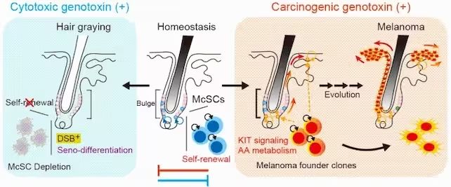
Under cytotoxic genotoxin exposure, such as X-ray irradiation, McSC self-renewal is impaired, leading to depletion and hair graying. In homeostasis, McSCs maintain self-renewal and pigment balance. Carcinogenic genotoxins, however, promote KIT signaling and alter arachidonic acid metabolism, triggering melanoma. (University of Tokyo)
Why this matters for skin aging and melanoma
The study reframes hair graying and melanoma as alternative outcomes of how stem cells handle stress. If a stem cell population chooses exhaustion via seno-differentiation, the tissue loses pigment but may gain protection against tumor initiation. If the microenvironment instead supplies survival signals, damaged stem cells may expand, raising cancer risk.
Importantly, the researchers are careful: gray hair is not itself a proven shield against cancer in humans. The work uses mouse models to reveal molecular circuits and plausible mechanisms. Confirming the same dynamics in human skin will require further studies that examine human McSC behavior, environmental exposures, and niche signaling in relation to both pigment loss and melanoma incidence.
Experimental details and molecular players
The team combined gene-expression profiling, targeted DNA-damaging treatments, and cellular lineage tracing in mice. They distinguish two broad damage contexts: cytotoxic genotoxins like ionizing radiation, which prompt p53-p21 activation and seno-differentiation; and carcinogenic exposures like UVB and DMBA, which engage growth-supporting niche factors and block the differentiation fail-safe.
Key molecules highlighted include p53 and p21 (damage sensors and cell-cycle regulators), SCF (stem cell factor) and KIT signaling (niche-derived survival cues), plus changes in arachidonic acid metabolism that appear tied to tumor-promoting inflammation. These pathways link the molecular response to observable outcomes: gray hair versus melanoma development.
Implications for prevention and therapy
Understanding the signals that tilt McSCs toward exhaustion or expansion opens potential interventions. Could modulating SCF/KIT signaling or the inflammatory milieu nudge damaged stem cells toward safe elimination? Might biomarkers associated with niche activity help identify skin at higher risk of melanoma even before tumors appear?
Translational prospects are speculative at this stage, but the conceptual advance is clear: tissue aging and cancer are not independent phenomena; they can be divergent consequences of how stem cells respond to genotoxic stress.
Expert Insight
Dr. Maya Reynolds, a dermatologist and stem-cell researcher (fictional), summarizes the study's practical angle: "This work connects visible aging — like graying — to deep molecular decision points in stem cells. If we can learn to read those decisions in human skin, we may gain new early-warning markers for melanoma risk and novel preventive strategies that act on the stem cell niche rather than the tumor itself."
Further research will need to validate these pathways in human tissues, map how lifetime exposures influence McSC fate, and explore whether manipulating niche signals can lower melanoma incidence without harming normal tissue maintenance.

“My work centers on sustainability, energy, and environmental science — examining how innovation can lead to a greener future.”

Discussion
Leave a Comment