A neurologist peers into stained tissue and sees something unsettling: immune cells swollen with bright, fatty droplets, clustered at the edges of damaged nerve fibers. These are not the usual inflammatory suspects. They are microglia, the brain’s cleanup crew, turned into fat-saturated, so-called foamy cells.
When cleanup becomes a problem
Multiple sclerosis can look like many diseases at once. For some people, it advances slowly, allowing decades of relatively normal life. For others, it progresses quickly, stripping away mobility and function in short order. New research from the Netherlands points to one possible reason why those paths diverge: microglia overloaded with lipids may not just be bystanders. They could be active drivers of the most aggressive disease courses.
The study examined post-mortem brain tissue from 28 people with secondary progressive multiple sclerosis and compared it with samples from 10 people without the disease. Using layered profiling methods that combine protein mapping, fat analysis, and gene activity measures, the team created an integrated picture of cells within MS lesions, where the myelin sheath that insulates nerve fibers has been attacked.

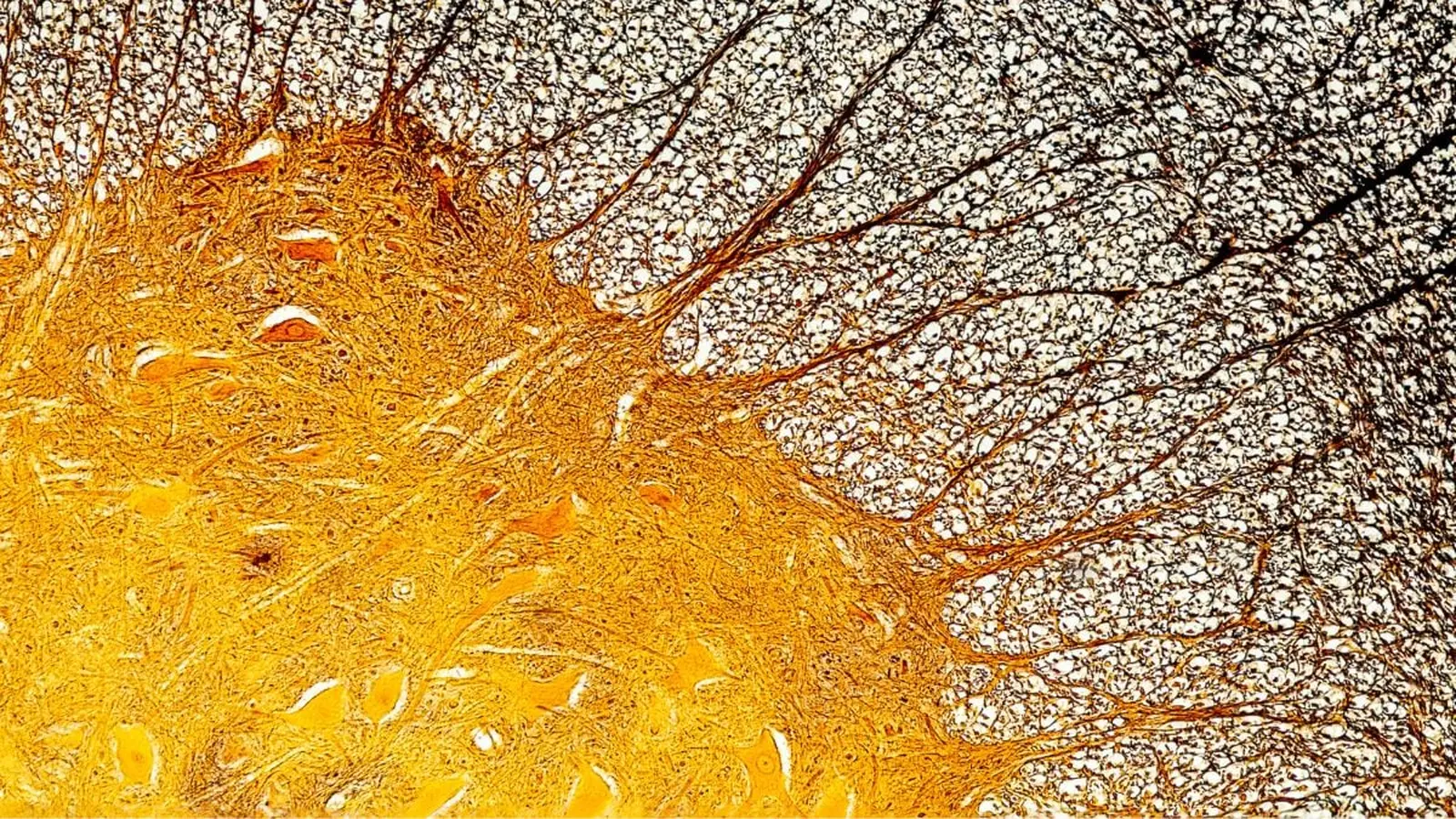
Images of MS lesions with foamy microglia. Red arrows indicated fat-filled pockets of increasing size (left to right) inside the immune cells.
Their finding: brains from patients with faster, more severe progression carried more of these fat-laden microglia. But quantity alone was not the whole story. The foamy microglia displayed a distinct molecular profile: different enzymes, altered protein signatures, and metabolic pathways skewed toward lipid handling rather than repair.
What the data suggest about mechanism
At a glance, microglia arrive at injury sites to clear debris and help rebuild. In MS lesions the debris includes myelin, the fatty insulation that nerve cells rely on to transmit signals. The new work proposes a tipping point. Microglia that ingest myelin begin to accumulate lipid droplets. Overloaded, their functional balance shifts. Instead of promoting resolution and tissue healing, they secrete factors that perpetuate inflammation and impede repair.
"We see these cells trying to clean up, but becoming saturated with fat," says Daan van der Vliet of Leiden University, the study’s lead author. "Once they are overloaded, their capacity to support repair declines and they may contribute to a harmful environment around lesions."
The team did not stop at human tissue. In a mouse model that mimics features of MS, researchers targeted one of the enzymes that was especially active in foamy microglia. When that enzyme was blocked, lesion healing improved. That intervention offers a direct, testable link between altered microglial lipid metabolism and worse tissue outcomes.
Mouse work and a potential druggable target
- The animal experiments showed better myelin repair when the enzyme activity was reduced.
- That suggests fat-processing pathways inside microglia might be amenable to therapeutic modulation.

Severe MS was associated with fat-laden, 'foamy' microglia.
Implications for diagnosis and treatment
Two implications emerge. First, if foamy microglia signal or cause faster decline, they could become a biomarker for aggressive MS. The researchers detected lipids linked to these cells in cerebrospinal fluid, raising the possibility of an assay that identifies high-risk patients earlier in the disease course.
Second, therapies that shift microglial metabolism away from harmful lipid accumulation, or that restore their repair functions, might slow progression. That idea complements other therapeutic strategies in MS that focus on suppressing immune attack. Here, the angle is metabolic: change how resident immune cells handle fats, and you may change their behavior toward tissue preservation.
Clinical translation remains a road with many turns. What works in mice often fails in people. The pattern of lipid handling in human microglia is complex, and interfering with metabolism has systemic effects. Still, the study supplies a plausible, testable mechanism that could explain why some forms of MS are so relentlessly damaging.

Expert Insight
"This research reframes part of the MS problem as a metabolic one," says Dr. Maya Singh, a neurologist and MS researcher at University College London. "If microglia switch from helpful cleaners to lipid-overloaded contributors to inflammation, then we need therapies that restore their balance. That could be a new avenue alongside immune modulation and neuroprotection."
The potential for cerebrospinal fluid biomarkers is particularly enticing. A simple test that flags patients at risk for rapid progression would change clinical decision-making. It could guide the timing and aggressiveness of treatment, focus monitoring, and even stratify patients for clinical trials of metabolism-targeting drugs.
Conclusion
The discovery of foamy, fat-saturated microglia in severe multiple sclerosis offers both explanation and hope. It explains a plausible biological pathway to faster decline, and it highlights fresh targets for intervention: enzymes and pathways that govern lipid processing in brain-resident immune cells. Moving from tissue maps to approved treatments will take time, and careful clinical studies will be essential. Still, this work opens a new line of inquiry in a disease that has long resisted a full explanation for its variability.
Related questions remain. How early do foamy microglia appear? Are they a cause or a consequence in every case? Could lifestyle factors or existing drugs influence microglial lipid metabolism? Answering these will require longitudinal studies, improved biomarkers, and eventually clinical trials.
For patients and clinicians, the message is pragmatic: researchers have identified a cellular change that correlates with worse outcomes and that can be manipulated experimentally. That is a clear step toward precision approaches in MS care, tailoring interventions to the biological features that drive each patient’s disease.

“The cosmos has always fascinated me. I write about space missions, astronomy, and the technologies pushing humanity beyond Earth.”

Discussion
Leave a Comment