Imagine standing at the threshold of a tiny workshop where molecular machines clank, rearrange, and respond. You cannot touch them. You cannot tag them. Yet the action is there, unfolding in real time. That is the new promise from a team at Stanford, who have stitched together two imaging traditions to reveal living cells at a level of detail not previously available without fluorescent labels.
Seeing cells without labels
For decades, fluorescence microscopy has been the workhorse of cell biology. Tag a protein, watch it move. The trade off is clear. Labels give specificity, but they also limit what you can see at once and how long you can watch it. Fluorescent dyes bleach. Genetic tags can perturb behavior. What if you could observe many structures simultaneously, without adding anything to the cell?
Enter Interferometric Image Scanning Microscopy, or iISM. The technique captures native scattering from cellular components and enhances that faint light through interferometry and a multi-element detector. The upshot is a label-free view that reaches about 120 nanometers in resolution, a scale where membrane subdomains, small organelles, and large protein complexes become visible in their natural context. That level of clarity had previously required fluorescence or very specialized instruments.
W.E. Moerner, a Nobel Prize recipient for his work in super-resolution fluorescence methods, and postdoctoral researcher Michelle Kueppers led the effort. They present the work in the journal Light: Science and Applications. Their claim is not triumphalist. iISM is complementary. It does not seek to replace fluorescence. Instead it offers a different set of strengths: context, longevity, and gentler illumination.

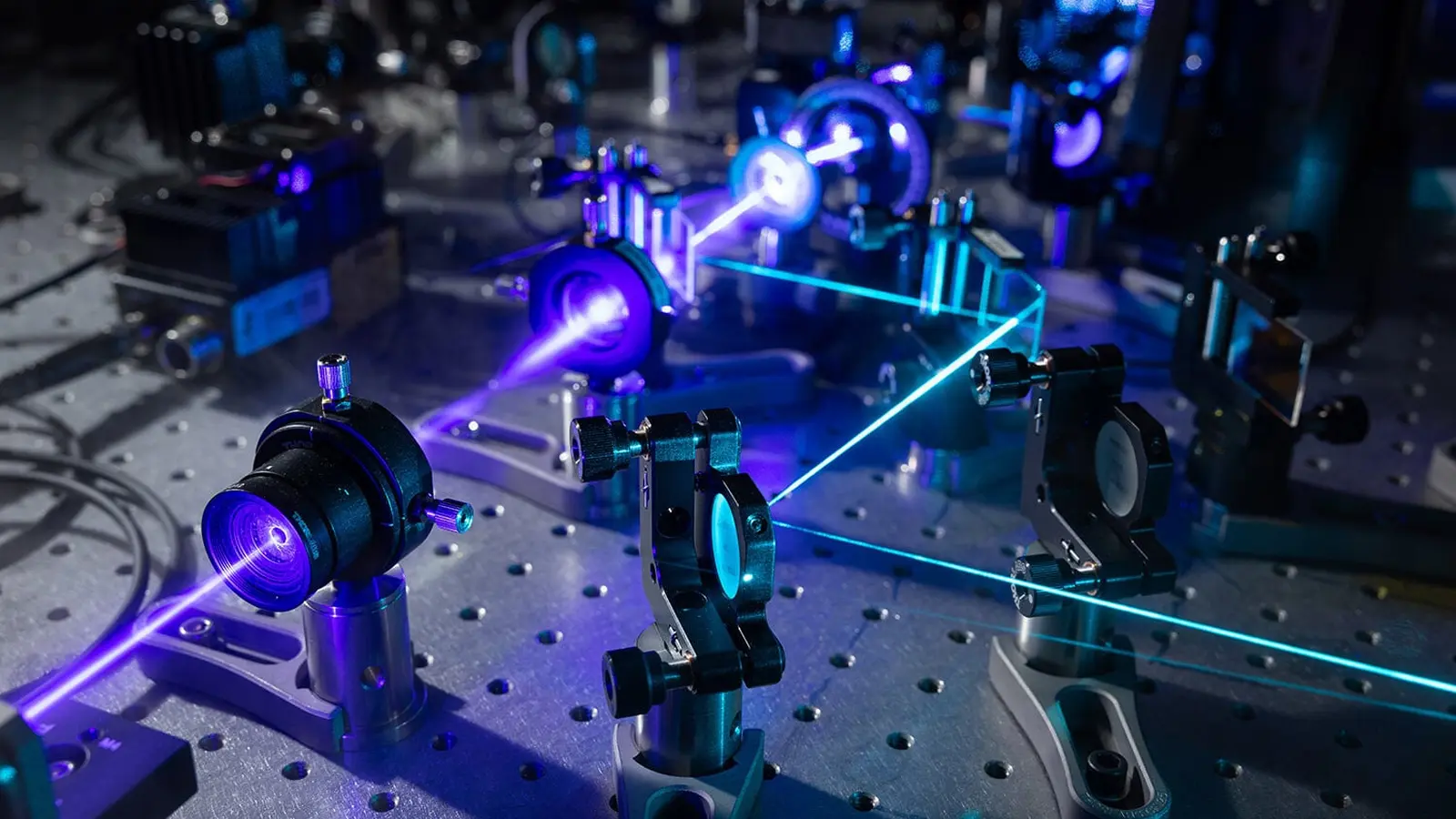
Michelle Kueppers and W.E. Moerner.
How iISM blends two traditions
At its core, iISM combines interferometric scattering microscopy with an adapted form of image scanning using camera arrays. Both pieces are established, but together they produce something new.
Interferometric scattering relies on a simple physical fact. When light encounters a small object, part of it scatters. That scattered light carries information about the object but is often extremely weak. Interferometry boosts the measurable signal by mixing the scattered wave with a reference beam, turning tiny phase and amplitude differences into detectable variations. This is similar to how radio receivers combine signals to improve reception.
Conventional confocal microscopes solve spatial blur by rejecting out-of-focus light with a pinhole and collecting photons with a single detector. Modern image-scanning approaches replace that single detector with an array of pixels. Each pixel captures a slightly different view of the same small volume. With many viewpoints you can computationally reconstruct a sharper image. Think of it as having tens to hundreds of eyes on the same scene rather than two.
Stanford’s team implemented an array detector that gathers more light than a pinhole and then developed a pipeline to fuse those multiple perspectives. The result is a label-free image with enhanced contrast and improved axial precision. Crucially, iISM achieves this while using lower illumination power compared with other high-contrast, label-free methods. Lower power means less photodamage and longer observation windows for living cells.
That combination opens windows into dynamics that are difficult to follow with fluorescence. Researchers can track multiple unlabeled features concurrently and observe their responses to stimuli such as pathogens or drugs. Because no labels are introduced, the structures are seen in their native state.
Applications, collaborations, and near-term prospects
The immediate appeal of iISM is broad. Moerner and Kueppers are already leveraging the instrument in collaborative projects across Stanford. One team is watching interactions among plant cells, fungi, and bacteria in real time. Another is following the entry path of a cancer drug into cells. A third plans to record how red blood cells deform under malaria infection. These are practical, high-impact problems where label-free context matters.
Beyond those early projects, the technique could influence several areas of life science research. Study of pathogen entry, membrane remodeling, intracellular transport, and drug uptake all stand to gain. In drug development, for instance, iISM could reveal how a candidate molecule navigates cellular barriers without the confounding effects of fluorescent tags. In plant biology, it can show multicellular interactions at the interface of host and microbe.
There are trade offs. iISM does not yet match the molecular specificity that targeted fluorescence can provide. It cannot identify a single protein among thousands on its own. But Kueppers and Moerner envision a hybrid future where fluorescence provides molecular identity while iISM supplies label-free context and dynamic range. Together, those views would give researchers both the who and the how.
Expert Insight
"This is a pragmatic advance," says Dr. Elena Park, a cellular imaging specialist at a research institute unaffiliated with the work. "What matters is not just resolution but the ability to watch processes unfold without perturbing them. iISM reduces the observer effect. That can change experimental design and open questions we could not frame before."
Dr. Park adds a caution. "Adoption depends on ease of use and access. If the technology becomes available to more labs, it will accelerate discoveries. If it remains a niche setup, progress will be slower."
Moerner and Kueppers appear to be addressing that barrier. They are iterating on the instrument and pursuing collaborations that demonstrate the method across diverse biological systems. That outreach matters for translation from a single lab to a community tool.
The practical benefits are clear. Lower phototoxicity, extended live imaging, simultaneous visualization of multiple unlabeled structures, and compatibility with fast dynamics provide a useful set of capabilities for many experiments. For questions that demand molecular specificity, fluorescence remains indispensable. For questions that demand context and gentle observation, iISM is promising.
In short, iISM adds a new, pragmatic lens to the microscopy toolbox. It favors subtlety over spectacle. It reveals motion in situ, without painting the cell in fluorescent colors. For scientists who want to watch life happen rather than label it, that is a major step forward.

“My work centers on sustainability, energy, and environmental science — examining how innovation can lead to a greener future.”

Discussion
Leave a Comment