5 Minutes
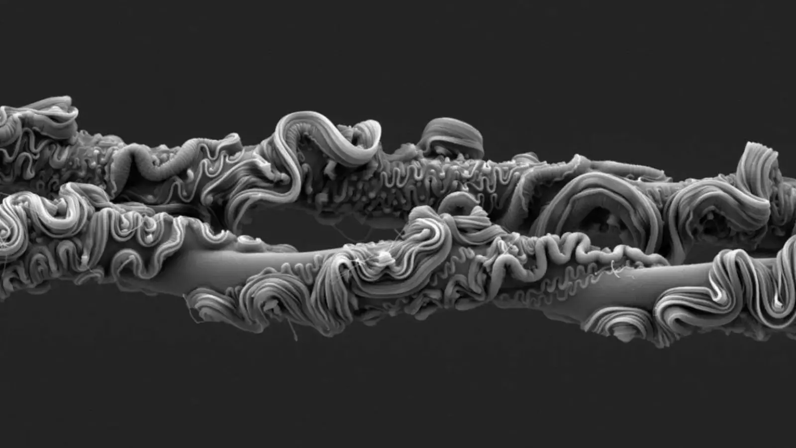
A striking electron-microscope image of spider silk has taken the top prize in the Royal Society’s 2025 photography competition. The winning photograph — a magnified study of the net-casting spider’s silk — reveals a composite structure that blends extreme elasticity with high tensile strength, offering both an arresting visual and new clues for biomaterials research.
A web engineered for hunting
The award-winning micrograph captures the silk from Asianopis subrufa, a species known as a net-casting or ambush spider. Unlike orb-weavers that passively wait for prey, net-casting spiders hold a small, rectangular net between their front legs and actively fling it over unsuspecting insects. Under the scanning electron microscope the silk appears as a nested architecture: an elastic inner core surrounded by multiple layers of stiffer filaments of varying diameters.

That combination of a stretchy core and layered, strong sheathing explains how the net can both deform rapidly to envelop prey and then resist tearing. The image not only highlights the biological ingenuity of a single predator’s toolkit, it also provides measurable microstructural detail that materials scientists can use to model load distribution, fatigue resistance and energy absorption in fiber composites.
Why the microstructure matters
Electron microscopy reveals features that are invisible at lower magnification — submicron fibrils, interfaces between layers and regions of variable density. These microstructures control mechanical properties such as Young’s modulus (stiffness), extensibility and toughness. For engineers trying to design flexible, tough fibers for applications from medical sutures to soft robotics, such natural blueprints are invaluable.
Biomimicry efforts have already borrowed spider silk concepts to create synthetic fibers with improved toughness. The Royal Society winner renews attention on net-casting spiders specifically: their silk must absorb and dissipate the kinetic energy of a mid-air capture, a functional challenge slightly different from the static load-bearing problems orb webs solve.
Other notable winners and global scenes

Photographer: Peter Hudson
In the animal behavior category, Peter Hudson captured two male greater prairie chickens (Tympanuchus cupido) in a dramatic lekking display. The photograph freezes the moment of aerial leaps and shoves used to assert dominance and attract mates — a vivid example of how behavior photography can illuminate mating strategies and territorial displays.

Photographer: Filippo Carugati
Filippo Carugati’s ecology entry shows tadpoles suspended in a gelatinous matrix on a tree trunk in Madagascar, likely belonging to Guibemantis liber. These field images document early life stages and microhabitats that are critical for conservation planning in biodiversity hotspots.
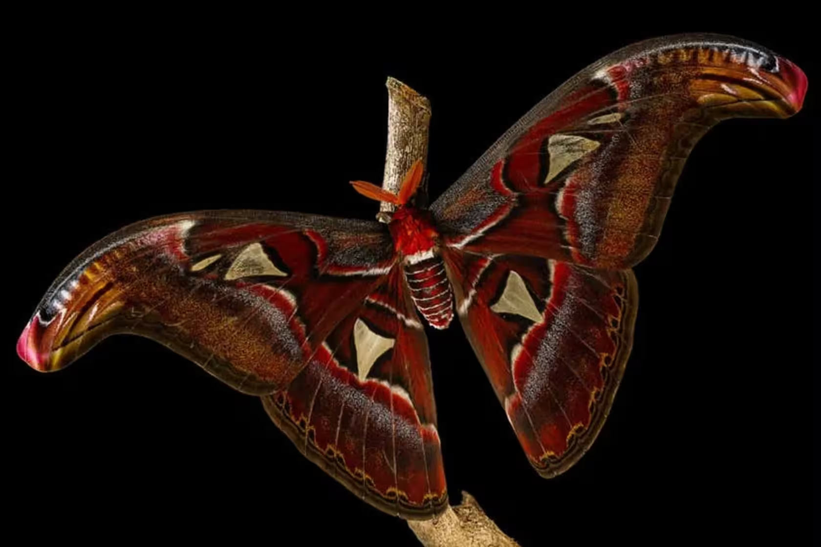
Photographer: Irina Petrova Adamatzky
Irina Petrova Adamatzky earned second place in animal behavior with an image of the Atlas moth (Attacus atlas) showcasing its snake-head wing tips — a classic example of morphological mimicry that reduces predation by birds.

Photographer: Felipe Rios Silva
In Earth and climate science, Felipe Rios Silva’s photo documents coastal stratocumulus clouds over the Atacama Desert, highlighting ongoing research into fog-harvesting techniques that convert marine moisture into potable water for arid communities.

Photographer: Aman Chokshi
Aman Chokshi captured the Antarctic sunrise after six months of polar night, producing a 360° panorama transformed into a stereographic “little planet.” The image, edged with green and purple aurora and crowned by the Milky Way, required extreme cold-weather camera preparation and patience in -70°C winds.
Scientific and technological implications
High-resolution images like these serve two purposes: they document natural phenomena with scientific precision and they catalyze interdisciplinary research. Materials scientists gain quantitative patterns for fiber design; ecologists and conservationists obtain visual records of rare behaviors and life stages; atmospheric scientists can use photographic evidence to communicate climate-related research to the public and policymakers.
Expert Insight
“This micrograph is more than a beautiful picture,” says Dr. Lena Ortiz, a materials scientist specializing in bioinspired fibers. “It provides a precise morphological map that lets us test hypotheses about how layered microstructures tune elasticity and strength. Translating those patterns into synthetic polymers could lead to lighter, tougher textiles and better-performing medical sutures.”
Images from international competitions like the Royal Society’s highlight how microscopy, fieldwork and creative vision combine to advance scientific understanding and inspire technology. From tiny silk fibrils to polar panoramas, each photograph is a data-rich story about how nature solves engineering problems.


Comments
atomwave
Cool photo, but is the sample prep altering silk structure? SEM can be harsh, so curious about artifacts, anyone know prep details?
labcore
Wow, that spider-silk micrograph is insane! Nature 1, engineers 0. Wish we could 3D-print that layering… but how scalable is it, really?

Leave a Comment