

Imagine a detective story in which the culprit leaves fingerprints far from the crime scene. In a new twist on how Alzheimer’s and related tau disorders unfold, researchers traced a damaging immune response back to the body’s lymph nodes rather than to the brain itself. The result challenges a long-held assumption that toxic protein clumps must be removed from the brain to prevent neuronal loss and cognitive decline.
Scientists traced a damaging immune response in tau-related neurodegeneration to signals originating in the body’s lymph nodes. In mice, disrupting this pathway protected neurons and cognition even though toxic tau tangles remained in the brain.
Looking past the blood-brain barrier
The blood-brain barrier usually stands between potential therapies and the neurons they aim to protect. It is a fortress of tightly regulated endothelial cells that limits drug entry into the central nervous system. Getting a treatment across that barrier is a major technical and financial hurdle. But what if you did not have to cross it at all?
Researchers at Washington University School of Medicine in St. Louis discovered that interrupting immune instructions originating outside the brain shields neurons from the cascade of damage linked to tau protein accumulation. The finding could reframe drug strategies: instead of designing molecules that penetrate the brain, scientists might target immune interactions in peripheral tissues where they are more accessible.
David M. Holtzman, MD, the study’s senior author, described the idea bluntly. Manipulating T cells has been routine in other fields of medicine. If those manipulations can be redirected to protect the brain indirectly, the therapeutic landscape changes considerably.
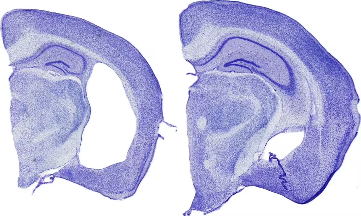
WashU Medicine researchers have found that blocking immune cells in the body’s lymph nodes significantly reduces Alzheimer’s-like neurodegeneration in mice. Shown are a mouse brain with tissue damage typical of Alzheimer’s-like neurodegeneration (left) and a brain in which the harmful immune response was prevented (right).
How immune cells deliver the damage
Brains affected by Alzheimer’s and similar disorders harbor more T cells than healthy brains. T cells are the adaptive immune system’s sentries. They usually protect against infection and cancer. In mouse models that develop tau tangles, earlier work from the same group showed that removing brain T cells reduced neuronal loss. That raised a fresh question: where do these T cells come from, and who tells them to attack?
The team zeroed in on classical dendritic cells type 1, or cDC1. Dendritic cells present molecular “wanted posters” to T cells, teaching them what to target. Only a handful of cDC1 reside inside the brain. The puzzling observation was that those brain-resident dendritic cells did not appear to be the teachers behind the T cell response that follows tau accumulation.
To test the origin story, researchers removed dendritic cells from lymph nodes and other peripheral sites in mice genetically programmed to develop tau tangles and neurodegeneration. Without those dendritic cells, the characteristic rise in brain-infiltrating T cells, especially CD8 T cells, never occurred. Equally striking, the mice were spared the typical brain tissue damage and showed preserved cognitive performance even though the tau tangles remained at similar levels.
That disconnect is important. It suggests that tau deposits and the immune-mediated neuronal loss are separable phenomena in these models. If immune activation is the amplifier that turns protein accumulation into progressive degeneration, then dialing down that immune signal could slow or prevent decline without eliminating tau itself.

The study appears in Nature Neuroscience. The authors caution that these results are in mice and that translating the approach to humans will require careful work to identify the precise molecular signals that draw T cells to the brain.
What triggers the peripheral immune response?
One plausible hypothesis is that tau-associated damage releases cellular debris or altered proteins that drain to cervical lymph nodes in the neck. There, dendritic cells could interpret those fragments as foreign or dangerous and prime T cells to respond. Those T cells then home back to the brain and, in some cases, drive neuronal injury.
At present the initiating signal remains unknown. Pinpointing it is a top priority because it offers a potential point-of-intervention that might prevent the misguided immune education before it sends destructive troops into the brain.
Expert Insight
"This study reframes where we look for therapeutic targets," said Dr. Maya Singh, a neurologist and immunology researcher unaffiliated with the study. "Targeting peripheral antigen presentation is attractive because it builds on decades of immunotherapy experience in other diseases. But we must be cautious; immune modulation has consequences elsewhere in the body, so any translation to patients will require precision."
Her remark highlights a practical tension: existing immune-modulating drugs could be repurposed, shortening the path to clinical testing, yet off-target effects or systemic immune suppression would be unacceptable in older adults at risk of infection.
Implications and next steps
The team is now testing whether interrupting dendritic cell function later in life, around the onset of tau pathology, can reproduce the protective effect seen when the manipulation begins at birth. They are also searching for the molecular breadcrumb trail that guides T cells to the central nervous system.
If the same mechanism operates in humans, the payoff would be substantial. Therapies could be developed to block the peripheral immune signal with existing classes of drugs that do not need to cross the blood-brain barrier, potentially lowering development cost and complexity.
At the same time, the research underscores the complexity of neurodegenerative disease. Protein aggregates, immune responses, vascular factors and glial cells all interact. The new work does not claim that tau tangles are harmless; rather, it exposes a secondary axis that converts proteinopathy into clinical decline.
Conclusion
This study opens a new chapter in how scientists think about Alzheimer’s and related tauopathies. By following immune traffic to its source, researchers found that lymph nodes and peripheral dendritic cells can orchestrate a brain-directed attack. The discovery shifts part of the therapeutic focus away from the brain and toward the immune system’s peripheral hubs. That shift could simplify drug delivery and reveal novel, more accessible targets for slowing or preventing neurodegeneration.

“My work centers on sustainability, energy, and environmental science — examining how innovation can lead to a greener future.”

Discussion
Leave a Comment
Comments (3)
My dad had Alz, so this hits different. If we can block peripheral immune signals without nuking immunity, sign me up. careful tho, no blunt hacks pls
Is this even true? Mouse models often mislead, curious what actually draws T cells to the neck nodes. need human data, pronto
Wow, lymph nodes sending T cells to wreck the brain? mind blown… if this holds in humans, whole approach flips. wild