
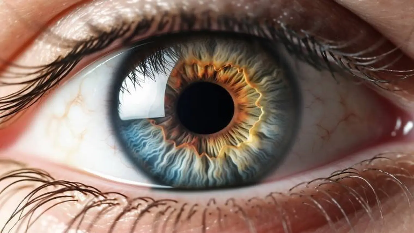
Imagine a clear cornea slowly overtaken by a creeping film, vision shrinking as healthy tissue is replaced. That is the daily reality for people with aniridia-related keratopathy, a rare genetic disorder in which the eye's limbal stem cells fail and conjunctival tissue spreads across the cornea. In a small but striking first-in-human study, researchers at Moorfields Eye Hospital and University College London used a collagen scaffold to deliver two types of stem cells directly to the damaged ocular surface—and patients saw measurable improvement.
Aniridia often traces back to mutations in the PAX6 gene, a key regulator of eye development. When limbal stem cells at the cornea's edge are depleted, the conjunctiva moves inward, the transparent cornea becomes opaque, and a progressively thickened surface robs people of sight. The condition is rare, affecting roughly one in 40,000 to one in 100,000 people, and until now has been difficult to treat effectively.
The trial enrolled nine patients. Each received the experimental treatment in one eye; the fellow eye served as a built-in control. The therapy combines a thin collagen scaffold with two cellular components: limbal epithelial stem cells, which re-seed the corneal surface, and stromal stem cells, which support tissue architecture and healing. The study, published in JAMA Ophthalmology, reports improvements in both ocular surface health and vision after implantation.
Clinical scores moved in a meaningful direction. At baseline the average ocular surface score, a metric that grades disease severity, sat in the moderate range for treated eyes. After three months the treated eyes improved to a mild score on average, and some benefit persisted at 12 months. Vision, measured with the ETDRS chart, increased by an average of 24 letters—roughly equivalent to nearly five lines on a standard eye chart.

Why this matters for corneal medicine
Why does delivering two cell types matter? One cell type restores the epithelial layer, the other helps rebuild the stromal environment that supports healthy growth. Together, they create a microenvironment more like the native limbus than a single-cell therapy could achieve. That combined approach appears to slow or reverse the encroachment of conjunctiva and to clarify the corneal surface enough for measurable vision gains.
Professor Sajjad Ahmad, consultant ophthalmic surgeon at Moorfields and professor of corneal regeneration at UCL, described the work as the first use of a collagen scaffold containing two stem cell types for an optical purpose, and said the team hopes to advance to a larger trial with a view to NHS adoption. The language is cautious, as it should be: nine patients is a small cohort, and longer follow-up will be needed to confirm durability and safety.
Still, this trial opens practical possibilities. A scaffold-based delivery system makes the therapy more controllable in the operating room and may be adaptable for other corneal surface disorders that share the same biomechanical problems. For patients with progressive aniridia-related keratopathy, it offers the first credible route to restoring sight rather than merely managing symptoms.
There are clear next steps: expand the study population, refine the manufacturing process for the scaffold and cells, and monitor outcomes over several years. If replicated in larger trials, this technique could shift the standard of care for a group of disorders that have frustrated clinicians for decades.


“My work centers on sustainability, energy, and environmental science — examining how innovation can lead to a greener future.”

Discussion
Leave a Comment
Comments
No comments yet. Be the first.