
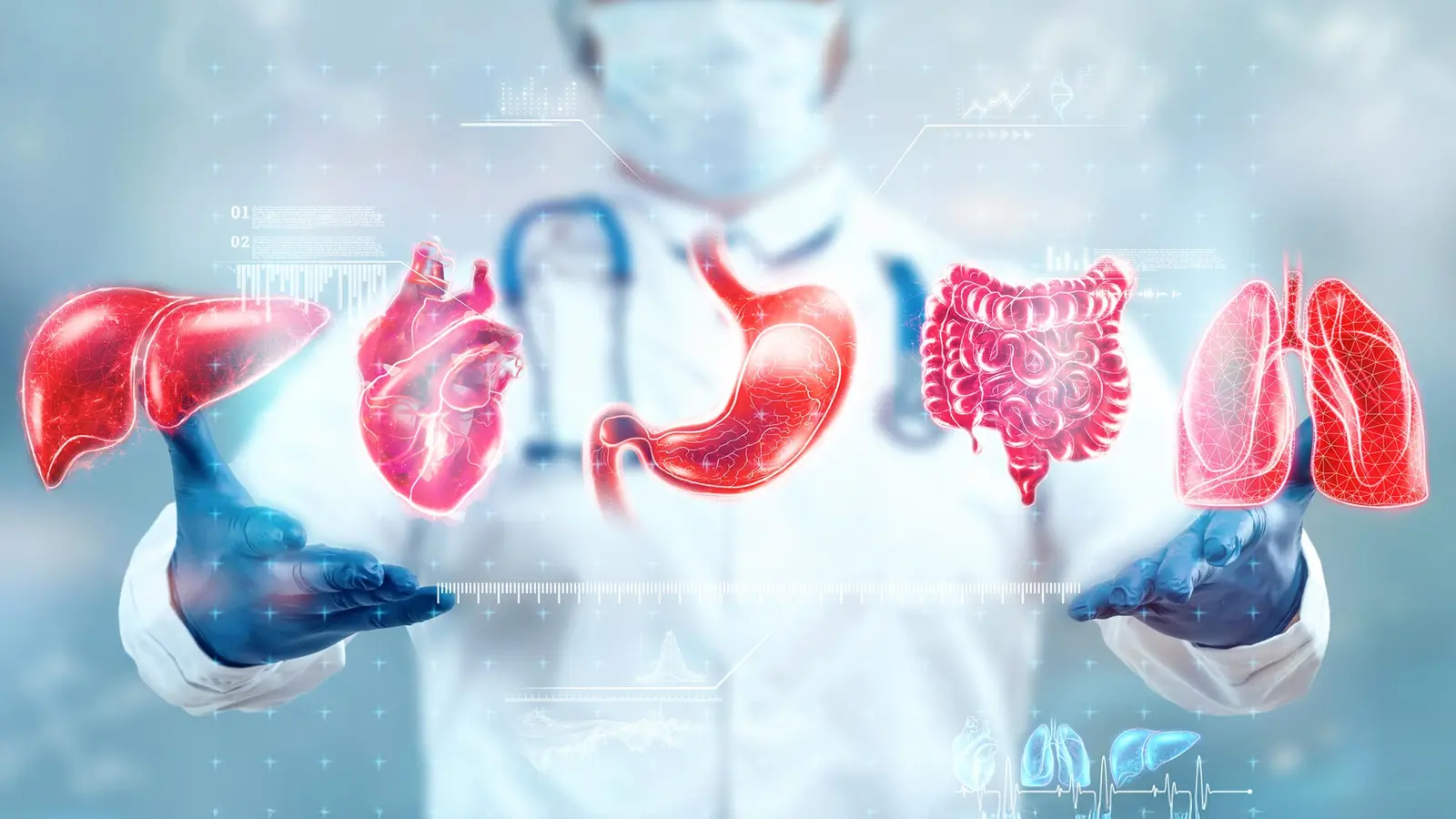
At a glance, a person and their organs tell two different stories. Chronological age sits on a birth certificate. Inside, tissues keep their own ledger, written in cells and scaffolding. Researchers have now taught artificial intelligence to read that ledger from routine histology images.
Training machines to read microscopic time
A team led by the CeMM Research Center for Molecular Medicine of the Austrian Academy of Sciences and the Ludwig Boltzmann Institute for Network Medicine at the University of Vienna asked a deceptively simple question: does the microscopic layout of a tissue carry a measurable imprint of age? The short answer is yes, and the method they used provides an unexpected window into how organs diverge from the clock printed on our passports.
Rather than focus only on molecular markers such as DNA methylation or gene expression, the scientists turned to histology, the study of tissues under the microscope. They used images from the Genotype-Tissue Expression Project, known as GTEx, which includes samples from 983 donors and spans about 40 tissue types. The dataset supplied 25,712 high-resolution images, which were subdivided into nearly half a billion image tiles and fed to advanced computer vision models.
The models were not explicitly trained to detect aging. Still, age emerged as the dominant factor shaping tissue appearance across the board. From those learned patterns the team built what they call tissue clocks, machine-learning predictors that estimate the biological age of a specific organ from its microscopic structure.

Histological image of a thyroid tissue sample obtained from the GTEx Portal.
Not all organs keep the same time
Prediction error across tissues averaged about 4.9 years, a level of precision that let the researchers compare organs within the same person. The results were striking. Some organs, including lung, kidney, pancreas, and adrenal gland, showed signs of accelerated structural aging relatively early, between the second and fourth decades of life. Others followed more complex, sometimes later trajectories. The uterus displayed a sharp architectural shift around menopause.
Why does this matter? Because pathology often begins as a change in tissue architecture. The tissue clocks were better at capturing organ-specific pathology than some DNA-based aging measures. Estimates of tissue age correlated with established hallmarks of aging, for example telomere shortening, plus the number of chronic conditions a person had. In practical terms, a heart that looks older than expected could flag damage or risk before symptoms arise.
"Our tissues retain a detailed record of aging," said André Rendeiro, the study's principal investigator. "Using histology and AI, we can detect patterns invisible to the naked eye and map the different ways aging unfolds across the body."

The authors of the study André Rendeiro, Iva Buljan, Ernesto Abila, and Yimin Zheng (f.l.t.r.)
Translating tissue signals into a blood test
Tissue biopsies supply rich information, yet they are not always practical. To bridge that gap the team matched tissue age gaps derived from histology with blood-based gene expression profiles from the same donors. The goal was to ask whether features of organ-specific aging could be read out indirectly, from a routine blood draw.
They built predictors that map signatures found in blood onto the tissue clock estimates. The resulting blood-based predictors flagged aging signals linked to several diseases. For example, Alzheimer disease showed the strongest aging signal in brain tissue, while Crohn disease associated with accelerated aging across gastrointestinal samples. Diabetes left a pronounced imprint on the pancreas. Other associations included vasculitis, cystic fibrosis, and stroke.
These findings suggest two things. First, tissue architecture encodes biologically meaningful changes that align with molecular aging and clinical disease. Second, those organ-specific signatures are not fully sequestered. Some portion of the signal leaks into circulation, where blood transcriptomes can act as a proxy, albeit an imperfect one.
Scientific context and methods in brief
The researchers applied deep learning to tile-level images, aggregated predictions across samples, and corrected for confounders such as sex and post-mortem interval. They compared tissue-clock outputs with telomere length, clinical diagnosis codes, and molecular readouts to validate biological relevance. This layering of pathology, gene expression, and clinical metadata is what gives the approach its power.
From a methodological perspective the study demonstrates the value of multi-modal data fusion. Histology provides spatial and structural context. Gene expression adds dynamic molecular state. Clinical records supply ground truth about disease. When combined, these layers improve sensitivity for organ-specific aging and pathology over single-modality assays.
Expert Insight
"This work reframes aging as a mosaic rather than a single dial," says Dr. Elena Marlowe, a geroscience researcher not connected to the study. "We often treat age as a uniform risk factor, but risk is organ-specific. Mapping which tissues are ahead or behind their expected schedule could change how we screen and intervene. The big challenge now is moving from retrospective tissue samples to prospective, minimally invasive monitoring."
Implications, translation, and future prospects
Several practical pathways emerge. One is improved disease surveillance. If blood tests can flag a pancreas that looks older than expected, clinicians might intensify monitoring for diabetes or intervene earlier. Another is drug development. Tissue clocks could serve as outcome measures for therapies intended to slow or reverse organ-specific aging.
However, important caveats remain. Most GTEx samples are post-mortem; therefore, caution is needed when extrapolating to living populations. Confounders such as medication history, environmental exposures, and demographic bias in the dataset deserve careful attention. Prospective cohorts, ideally with repeated sampling, are required to determine whether tissue-clock changes predict future disease and can be modified.
Ethical and clinical pathways must also be considered. If a blood test indicates accelerated aging in an organ, what follow-up is appropriate? How should risk be communicated? Which populations stand to benefit most from screening? These questions will determine how tissue clocks move from research to routine care.
Conclusion
The study offers a provocative new angle on aging: tissues keep structural memory that can be read by machines, and some of that memory is audible in blood. Organs do not age in unison. They have distinct tempos and susceptibilities, and recognizing those differences may enable earlier diagnosis, more targeted monitoring, and new endpoints for therapies aimed at healthy aging. The path from microscope to clinic will require validation, thoughtful translation, and careful ethical framing, but the map is beginning to take shape.

“The cosmos has always fascinated me. I write about space missions, astronomy, and the technologies pushing humanity beyond Earth.”

Discussion
Leave a Comment
Comments (3)
Seen tissue changes in reports at work, this could be a game changer for screening. but need longitudinal studies, and clear followup plans.
Promising, but GTEx is mostly post-mortem and biased. can blood transcriptomes really reflect organ architecture? sounds optimistic, cautious tho
whoa, organs aging at different paces? that's wild. a heart could look older long before pain shows... makes me curious, kinda scared lol