
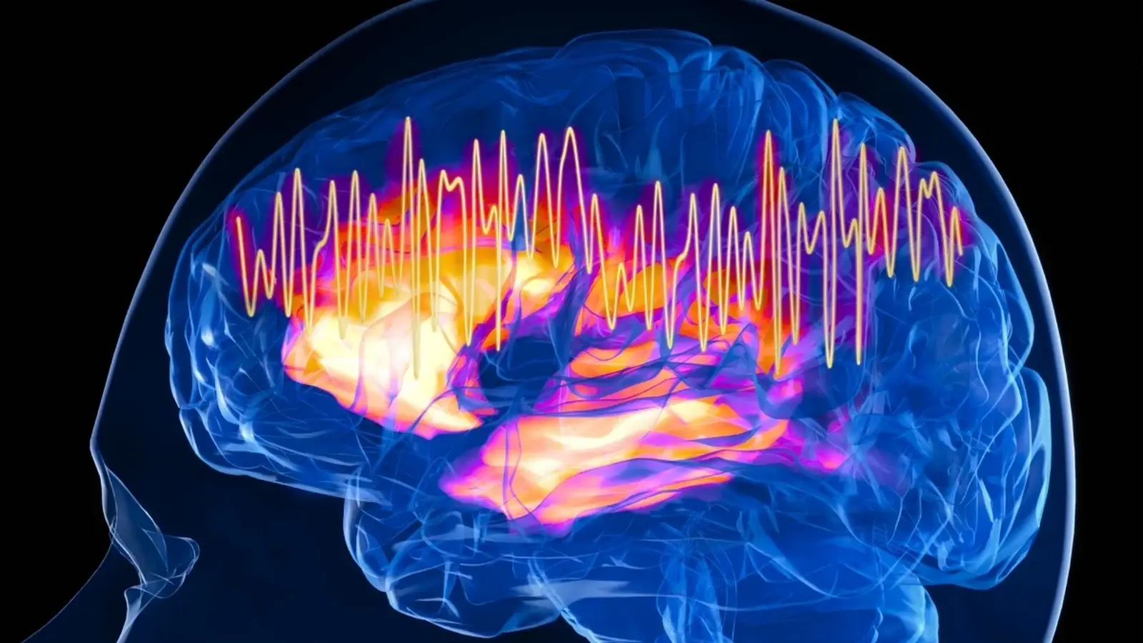
Attention can feel like a muscle you either have or do not. But what if that muscle is ancient, carved deep into the vertebrate brain long before humans showed up? Recent experiments at Johns Hopkins suggest that a tiny cluster of inhibitory neurons, preserved across birds, fish, and mammals, acts as a kind of focus switch in mice. When those cells are turned off, animals become distractible in a way that echoes symptoms of human attention disorders.
A locked-on stare, wired into the brain
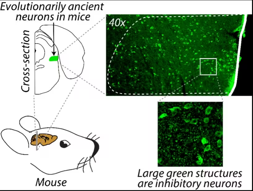
Not all attention lives in the cerebral cortex. For decades, neuroscientists have emphasized modern brain circuits, especially those shared by primates, as the seat of selective spatial attention. The new study redirects the spotlight to a much older structure. The neurons in question belong to the parabigemino lateral tegmental inhibitory complex, abbreviated PLTi. They release GABA, the central nervous system's main inhibitory neurotransmitter, and they modulate the superior colliculus, a midbrain hub that builds a spatial map of the world and helps orient perception toward important targets.
Why does this matter? Because the superior colliculus interacts with vision, hearing, and movement systems to guide where an animal looks and what it processes. Think of it as an internal stage manager, dimming the background and highlighting the actor we need to see. The PLTi applies a kind of selective blackout, muffling lesser signals so a single source can dominate processing.
The experiment that silenced focus
Researchers trained mice on a touchscreen task that required them to nose tap the correct image while ignoring irrelevant stimuli. With the PLTi intact, the animals performed reliably and earned rewards. Then the team used a viral tool to temporarily shut down the PLTi. The result was immediate and dramatic. Mice became prone to attending to faint distractors and were much less likely to pick the target location containing the useful information.
What the behavioral data showed
- Mice with active PLTi circuits ignored distractors and selected high priority targets.
- When PLTi neurons were inactivated, distractors pulled attention away, even when they were weak.
- Reactivating the PLTi restored focused behavior on the following day.
"Animals possess the remarkable ability to select and preferentially process the highest priority stimulus in space while ignoring distracting stimuli of lower priority," the authors wrote. Johns Hopkins neuroscientist Shreesh Mysore described the PLTi as an attentional selection engine. "The only thing impaired was the ability to take competing pieces of information, compare them, and pay attention to the location with the most important information," he said. His colleague Ninad Kothari summarized the behavioral change bluntly. "When we inactivate these neurons, the mice become hyper distractible."

Why this matters for human attention
Mice are not humans, and the authors acknowledge differences in brain structure and function. Still, the PLTi sits in a network that is highly conserved across vertebrates. Given that conservation, it is plausible that similar circuits exist in people and contribute to how we filter sensory inputs. The study also ties into a larger story about GABA signaling. Disruptions in inhibitory neurotransmission have been implicated in attention-deficit/hyperactivity disorder and other psychiatric conditions, so finding a discrete group of GABAergic neurons that gates spatial attention offers a new lead.
Practical implications are speculative at this point. Yet the experimental toolbox available in mice allowed scientists to silence a circuit and observe near-immediate behavioral consequences. Ethics committees would not permit that kind of intervention in humans. That constraint makes animal models indispensable for mapping cause and effect in complex brain functions.
Could this research inform treatments for ADHD or schizophrenia? Possibly, but cautiously. If homologous neurons or pathways are identified in the human brain, they could become targets for neuromodulation or pharmacology designed to restore inhibitory balance. Translational work will require imaging, postmortem studies, and careful physiological mapping in people and nonhuman primates.
Expert Insight
Dr. Laura Chen, a cognitive neuroscientist not involved with the study, commented on the findings. "What is striking is how an evolutionarily ancient node influences such a high-level behavior as selective attention. This challenges us to think beyond cortical architectures when we model attention. From a clinical perspective, pinpointing conserved inhibitory hubs opens new avenues for targeted research on attentional disorders."
The research, published in Nature Communications, reframes selective spatial attention as a distributed process that includes deep brainstem components as well as cortical networks. That distribution might explain why attention is robust in some contexts and fragile in others. When the PLTi functions correctly, distractions fade into the background. When it falters, even faint stimuli can pull the gaze away.
Conclusion

The discovery of PLTi neurons as key modulators of spatial attention in mice adds an important piece to the puzzle of how brains decide what to process and what to ignore. It highlights an ancient circuit with modern relevance, one that could bridge basic neuroscience and clinical research on attention disorders. For now, the study supplies a clear experimental demonstration: silence this small group of cells, and focus evaporates; switch them back on, and focus returns.

“My work centers on sustainability, energy, and environmental science — examining how innovation can lead to a greener future.”

Discussion
Leave a Comment
Comments (2)
hmm is this even true in humans? mice results nice but primate studies needed, fast translation sounds premature
wow didn't expect the brainstem to gate attention, wild… mice totally lose focus when those cells go quiet. kinda eerie but promising for ADHD work