
Imagine opening a box of memories and finding one corner a little emptier than it used to be. That is the unsettling image researchers are beginning to paint about a key memory structure in the aging brain and its link to depressive illness.
What the researchers revealed
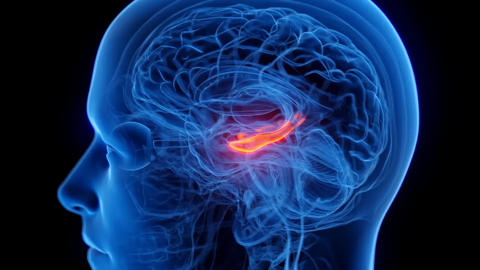
In an analysis of MRI and PET scans from more than 2,000 adults aged 50 to 90 who were cognitively unimpaired, scientists identified a specific hippocampal subregion that is smaller in people with a history of depression. The region is known as CA23DG, a composite area that groups together the CA2 and CA3 fields with the dentate gyrus. These subfields play distinct roles in forming and retrieving memories, pattern separation, and in guiding navigation through our environment.

People with depression show reduced volume inside a part of the brain that's essential for memory – the hippocampus.
After controlling for age, sex, body mass index and activity levels, CA23DG volume remained lower in participants who had experienced depression compared with those who had not. That relationship held even when the investigators accounted for canonical Alzheimer’s disease markers, such as brain amyloid and tau protein levels, and the presence of the APOE ε4 genetic variant. In short, the finding points to a hippocampal change that may be independent from the classical biological fingerprints of Alzheimer’s disease.
Not every hippocampal corner showed shrinkage. The CA1 subfield and the subiculum did not display the same consistent reduction in the baseline analysis. But the picture changed when antidepressant use entered the model: participants who reported taking antidepressant medication had smaller volumes not only in CA23DG but also in CA1. That was unexpected. Medication is supposed to relieve the harmful physiological effects of depression over time, not add to structural decline.

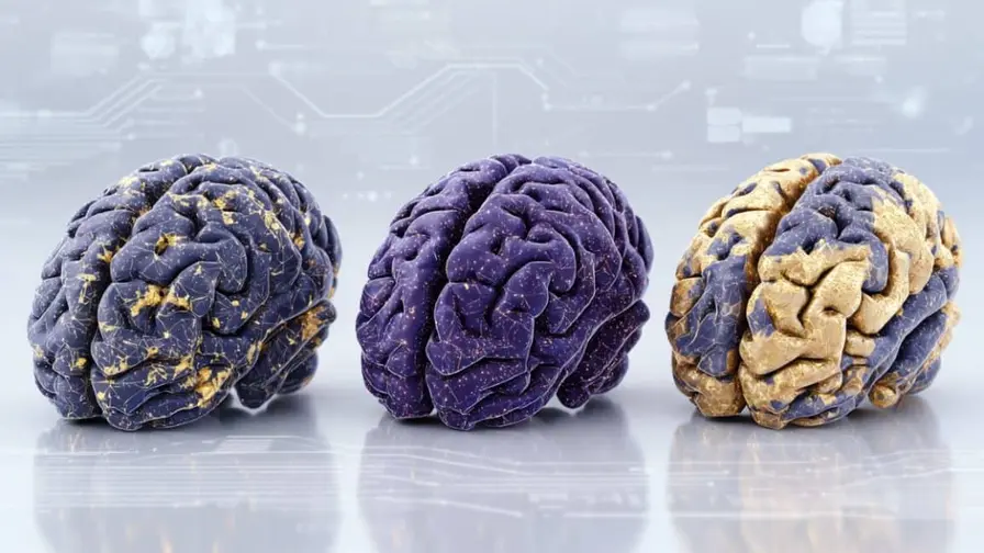
Comparison image showing what hippocampal atrophy can look like.
So what is driving these differences? The study’s authors suggest two nonexclusive possibilities. One, the structural change may reflect a distinct mechanism tied to depression itself, separate from Alzheimer’s biology. Two, medication exposure or the fact that people with more severe, longer-lasting depression are more likely to be prescribed antidepressants could be behind the association. The current dataset cannot definitively separate those explanations.
Implications and scientific context
We have long known epidemiologically that depression increases the risk for later cognitive decline and dementia, including Alzheimer’s disease. What has been murky is the biological route that links mood disorders and memory loss. The new finding narrows the search. It highlights CA23DG as a potential locus of vulnerability in older adults with depressive histories.
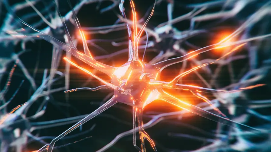
The dentate gyrus and CA3 are central to encoding new memories and distinguishing similar experiences. If those microcircuits are compromised, subtle memory deficits could follow. But causation is not established.
Duration appears to matter as much as severity. Participants who had experienced depression for longer periods—sometimes even with milder recent symptoms—showed a stronger association with reduced CA23DG volume than those with more acute or newly diagnosed episodes. That pattern hints that cumulative exposure to depressive episodes, stress hormones, or related inflammatory signals over years might be relevant.
At the same time, the researchers caution against interpreting these results as evidence that antidepressants cause hippocampal shrinkage. It may be that more severe or persistent illness both drives medication use and leaves a larger structural footprint in the brain. Longitudinal studies that scan people before and after treatment will be essential to untangle cause from correlation.
Expert Insight
'This study gives us a much more granular look at where depression and memory circuitry intersect,' says Dr. Elena Marquez, a cognitive neuroscientist at the University of Cambridge. 'The hippocampus is not a single entity. Finding a localized association in CA23DG is valuable because it directs follow-up work to specific cell types and pathways. Still, we need prospective imaging and careful tracking of medication histories to know whether the observed shrinkage is a marker of past stress, a medication signal, or some combination of both.'

People who experienced depression for longer showed a stronger association with the reduced CA23DG volume.
What comes next for research and care
Future studies will need several features: repeated imaging over years, detailed records of antidepressant type and dosage, objective measures of depressive symptom history, and markers of stress biology such as cortisol and inflammatory cytokines. Animal models that isolate specific subfields may reveal cellular mechanisms—whether it is loss of neurons, changes in synaptic density, reduced neurogenesis in the dentate gyrus, or other microstructural alterations.
Clinically, the immediate takeaway is caution and perspective. The new work does not justify stopping treatment. Antidepressants remain a mainstay for managing depression, and untreated depression itself carries risks for quality of life and possibly cognition. Patients should discuss concerns with their clinicians rather than make medication changes based on a single study.
Conclusion
The finding that a precise hippocampal subregion, CA23DG, may be smaller in older adults with depression adds a focused piece to a complex puzzle linking mood disorders and memory decline. It invites targeted follow-up, both to confirm whether this is a signature of depression, medication effects, or an interaction of factors, and to explore interventions that might protect vulnerable hippocampal circuits as people age.

“My work centers on sustainability, energy, and environmental science — examining how innovation can lead to a greener future.”

Discussion
Leave a Comment
Comments (2)
My dad had long bouts of depression and tiny memory slips. reading this makes sense, scary tho. Not saying stop meds, but worth more research, idk
Wait CA23DG? Sounds specific, but is this causal or just correlation... meds confuse things. Need scans before/after treatment, idk.