
Picture a mouse returning to a place that once shocked it. It freezes, alert and immobile, until something in its brain finally signals, You are safe now. That signal may involve a small set of neurons tuned to oxytocin, the hormone we usually associate with trust and bonding.
Where love hormones and danger assessments meet
Oxytocin has a reputation. It s the so-called love hormone, linked to pair-bonding, parental care, and social trust. But the molecule does more than glue social bonds together. New experiments in mice point to a tiny group of oxytocin receptor-expressing cells in the paraventricular thalamus, or PVT, that appear to influence both the desire to approach others and the ability to learn that a threat is gone. In short: social behavior and fear extinction converge in one neural neighborhood.
Researchers in Japan set out to map that neighborhood. They used genetically modified male mice to manipulate only the neurons that express oxytocin receptors in the PVT. By switching those cells on or off, the team probed how the animals behaved in social tests and in a classical fear-extinction paradigm.

What the experiments showed
Turning those neurons off reduced social approach. The mice were less interested in investigating unfamiliar conspecifics across two different behavioral assays, and the change could not be explained by sluggish movement or heightened baseline anxiety. But turning the cells on did not make mice suddenly gregarious. That asymmetric effect suggests the neurons are necessary for normal social engagement but not sufficient to boost it on command.
Fear extinction revealed another facet. The mice learned to associate a context with a mild shock. When returned repeatedly to that context without shocks, typical animals gradually stopped freezing. Mice with suppressed oxytocin-sensitive PVT neurons, however, kept freezing: they failed to update their memory to reflect safety. Activating the same PVT population reduced freezing early in extinction training, but the benefit faded; it did not persist to tests one day or one week later.
The specificity matters. Manipulating oxytocin-sensitive neurons in other brain regions did not alter sociability, demonstrating that oxytocin s effects depend strongly on circuit location. This regional nuance helps explain decades of mixed findings where oxytocin sometimes promotes approach and sometimes avoidance.
Human links and limitations
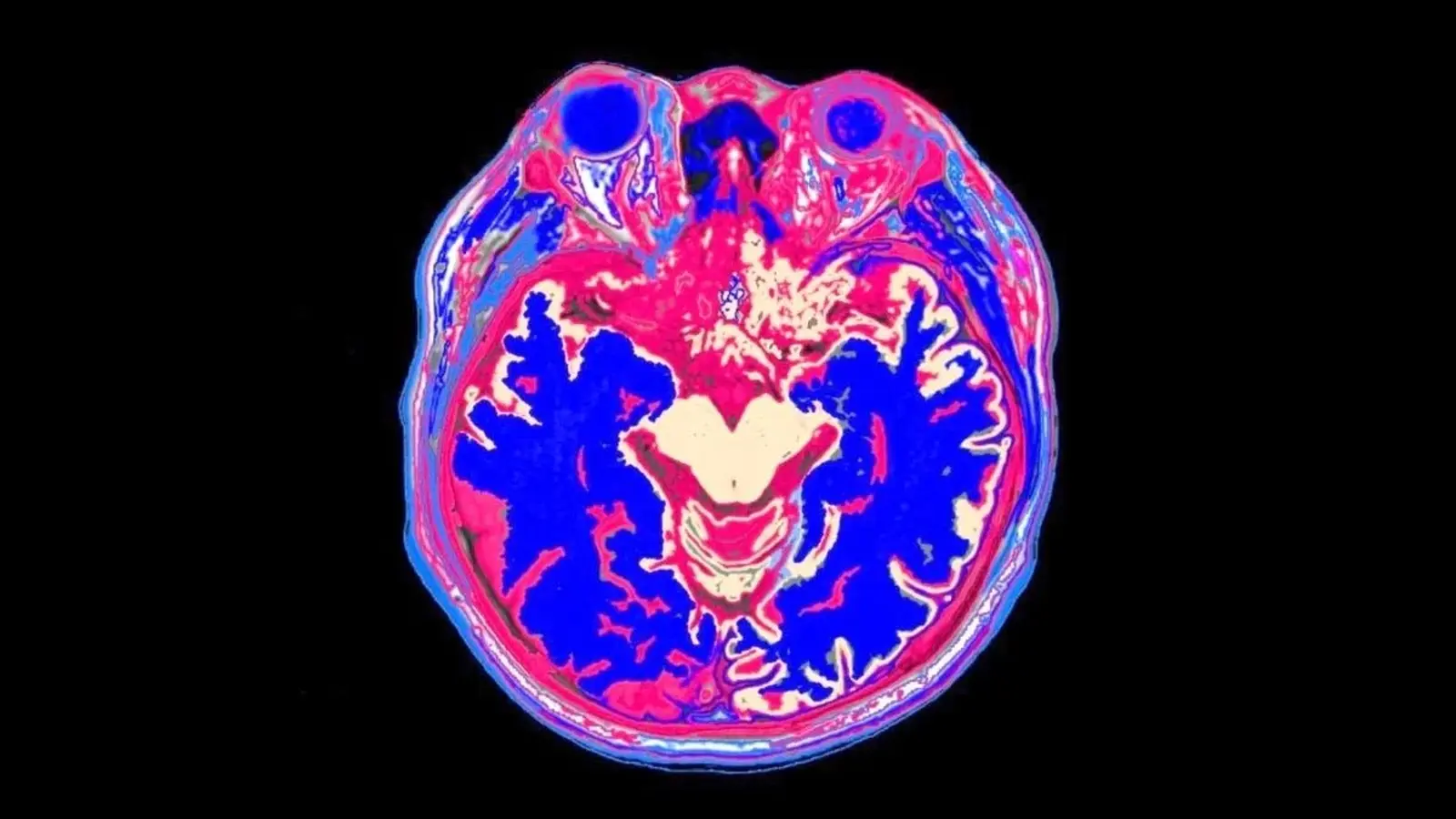
The team also ran a small, exploratory human study. They compared salivary oxytocin, MRI measurements of thalamic features, and behavioral assessments in 30 Japanese adolescents, 19 of whom had autism spectrum disorder. The results hinted at relationships between oxytocin, thalamus anatomy, and certain behavioral traits. But the sample was small and saliva measures are an imperfect proxy for what happens inside the brain. The animal work has its own caveats too: mostly male mice were used, and artificially activating receptor-expressing neurons does not replicate the brain s natural, timed release of oxytocin.

Why this matters for disorders of social behavior and fear regulation
Disruption of social approach and impaired fear extinction are features of conditions such as autism and anxiety disorders. If the PVT oxytocin-sensitive population plays similar roles in humans, that circuit might help explain overlapping symptoms. Importantly, the findings argue against one-size-fits-all treatments that simply increase oxytocin levels body-wide. Targeting specific circuits, or timing interventions to natural release patterns, could prove more effective.
Key caveats in plain terms
- The mouse manipulations are powerful but artificial; they do not mirror natural oxytocin signaling dynamics.
- Sex differences were not explored extensively; female responses could differ markedly.
- Small human sample and peripheral hormone measures limit translational claims.
Expert Insight
Dr. Lena Mori, a neurobiology researcher unaffiliated with the study, says: 'This work reframes oxytocin as a context-dependent modulator rather than a universal social enhancer. The elegant targeting of PVT cells shows that where oxytocin acts is as important as whether it is present. Translationally, that directs us to ask how timing, receptor location, and sex shape behavioral outcomes.'
Conclusion
The study adds a precise piece to a complex puzzle: oxytocin-sensitive neurons in the paraventricular thalamus help bridge social approach and the learning that a threat has passed. More work is required to map the circuitry in humans, to test female biology, and to determine whether manipulating these pathways could alleviate social or anxiety-related symptoms. For now, a clear message emerges: location matters. The same molecule can have very different effects depending on which neural circuit it touches.

The location of oxytocin action matters more than its mere presence.

“My work centers on sustainability, energy, and environmental science — examining how innovation can lead to a greener future.”

Discussion
Leave a Comment
Comments (2)
this is wild! tiny brain cells linking trust and fear, omg the therapy implications are huge... but still need more human trials, pronto
Wait so oxytocin isn't a universal 'love' fix? interesting but is this in females too? kinda worried about male-only bias.