

The skull may be doing more than protecting the brain. New human imaging and tissue data suggest that bone marrow inside the skull can light up with immune activity in people who live with long-term pain, hinting at a previously overlooked source of neuroinflammation.
A conduit no one expected
Back in 2018 researchers discovered tiny channels that connect the skull’s bone marrow to the membranes surrounding the brain. Think of them as little service tunnels for immune cells. That finding reframed how scientists imagine brain immunity: not only do immune cells travel from distant bone marrow, they may also use these local pathways to act fast and locally.
What happens when those local pipelines become chronically active? That is the question a team led by clinical imaging scientist Mehrbod Mohammadian at Harvard Medical School sought to answer in a study published in Science Translational Medicine.
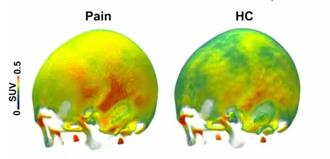
An averaged representation of TSPO PET signals recorded in the study, with yellow/orange/red colors corresponding to elevated levels of the protein in the chronic pain group (left) compared to the healthy controls (right).
Skull marrow and chronic pain
The researchers analyzed PET and MRI scans from 125 adults with persistent pain and compared them with scans from 22 healthy volunteers. The chronic pain cohort included 88 people with ongoing back pain and 37 with knee osteoarthritis. The imaging targeted translocator protein, or TSPO, a molecule that rises in many immune cells and serves as a marker of neuroimmune activation.
Scans showed elevated TSPO signals across large swaths of the skull in people with chronic pain. The increases were particularly strong in frontal and parietal skull regions. People with knee osteoarthritis displayed the most pronounced signals. Higher skull TSPO tended to align with self-reported pain intensity and how much pain interfered with daily life, and some analyses linked the signal to anxiety and depressive symptoms.
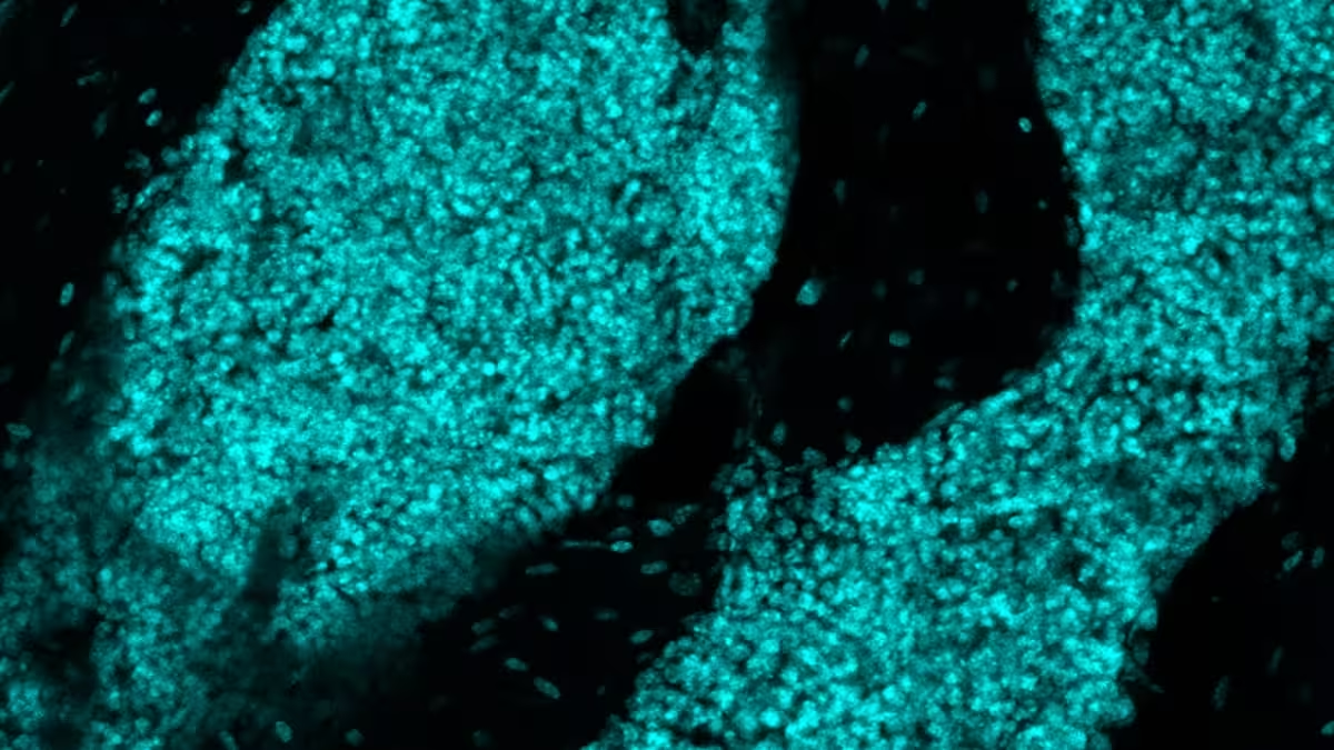
Recently discovered structures found in the skull bone marrow of mice.
Why TSPO matters
TSPO is abundant in myeloid immune cells that reside in bone marrow. When TSPO lights up on a PET scan it does not give a cellular play-by-play, but it reliably flags areas where immune activity is increased. In the context of this study, elevated skull TSPO is consistent with heightened immune engagement inside skull marrow.
From scans to tissue
Imaging was only one piece. To complement the in vivo findings, the team examined donated skull bone marrow samples from two 62-year-old men: one with a history of chronic pain and one without. The sample from the donor who reported chronic pain contained more than twice as many total cells as the control sample, and a larger share of those cells were TSPO-positive: 3,826 total cells with 82 percent TSPO-positive versus 1,616 total with 55 percent TSPO-positive in the control. While the authors caution that post-mortem tissue data are preliminary, the pattern echoes what the scans suggested.
Recently discovered structures found in the skull bone marrow of mice, as reported here. (Jang Hyun Park)
These convergent signals point to a hypothesis: skull marrow can mount immune responses that interact with the brain through skull-meninges channels, mobilizing myeloid cells and potentially driving local neuroinflammation that correlates with chronic pain.
- Possible mechanism: brain-originating immune signals may travel to skull marrow, activate local myeloid cells, and then feed back to brain tissues.
- Clinical correlation: higher skull TSPO links with greater pain and disrupted daily function.
- Open question: whether skull marrow activity causes pain, or reacts to it, remains unresolved.
Implications for therapy and research
This line of work opens a new therapeutic target: the skull itself. If immune activity in skull marrow contributes to chronic pain, then targeted interventions that calm or modulate that local immune compartment could provide relief. That might include drugs that alter myeloid cell behavior, targeted delivery across the skull, or neuromodulation approaches that influence skull-to-brain signaling.
But caution is required. The study is observational and associative. Imaging markers like TSPO tell us where immune activity is higher, not the causal chain. Experimental studies, longitudinal imaging, and carefully controlled interventions will be needed to determine whether tamping down skull marrow activity reduces pain.
Expert Insight
"These findings offer a fresh angle on chronic pain—one that moves the focus from the brain alone to its bony envelope," says Dr. Laura Baines, a clinical neurologist and pain researcher not involved with the study. "It suggests new experimental strategies: monitor skull marrow over time, test whether treatments that alter peripheral immunity shift skull signals, and then see whether those shifts predict symptom change. That would move us beyond correlation."
Conclusion
The skull is no longer merely armor. Imaging and tissue data converge to suggest that skull bone marrow is an active immunological hub that associates with chronic pain. Whether this activity is a driver or a passenger in the experience of long-term pain remains to be proven. Still, by spotlighting the skull marrow, researchers have opened a promising path: one that could reshape pain biology and eventually inspire treatments that target the brain’s most overlooked neighbor.

“The cosmos has always fascinated me. I write about space missions, astronomy, and the technologies pushing humanity beyond Earth.”

Discussion
Leave a Comment
Comments (2)
is TSPO specific enough tho? PET shows immune activity but doesn't say who's driving it, cause or reaction? quick thought
wow, skull marrow lighting up with immune activity... wild idea! could reshuffle how we think about chronic pain, but lots to prove.