
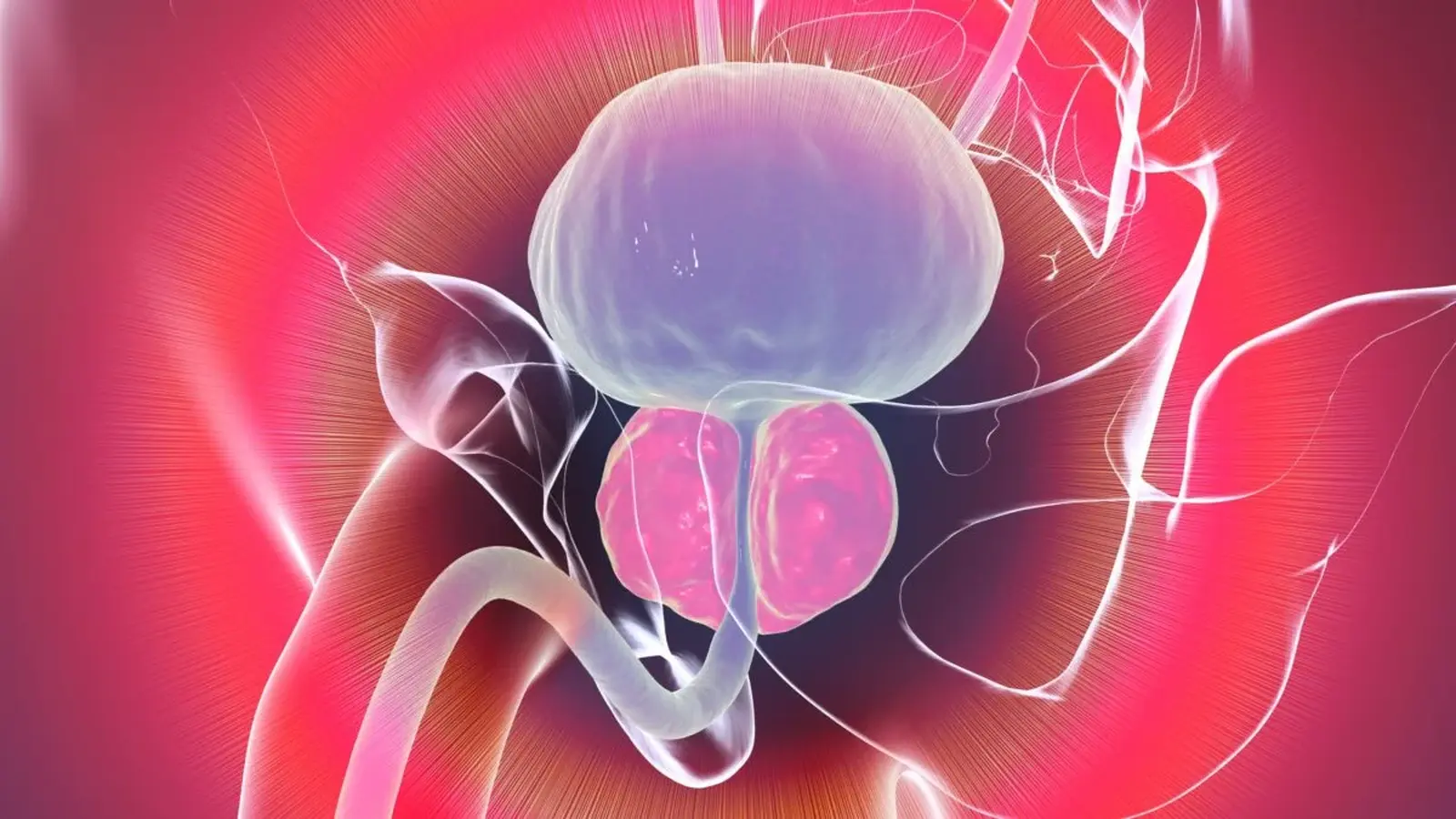
When the prostate begins to crowd the urethra, the result is not pain but a stubborn, urinary traffic jam. Men over 50 learn that routine quickly. For decades doctors have blamed hormones, inflammation, or simple wear and tear. But a recent study from Johns Hopkins points the finger at a specific population of cells that cluster around the urethra, and that could finally clarify why the gland enlarges with age.
A hidden population next to the urethra
Researchers dug into nearly 130,000 individual prostate cells using single-cell RNA sequencing. The tissue came from 10 men undergoing prostate cancer surgery, although the samples examined were noncancerous. Instead of treating the gland as uniform, the team sampled the three major anatomical zones and mapped which genes were turned on in each cell.
What they found was a distinct subtype of basal epithelial cell, rare outside one region but abundant in the part of the prostate that wraps around the urethra. The authors call these cells WIF1 positive or WIF1+ basal cells. On a gene-expression level they look different from other basal cells. Some of their signature genes are sensitive to androgens such as testosterone and feed into signaling circuits known to shape prostate development and growth.
Why does that matter? Because benign prostatic hyperplasia, commonly abbreviated BPH, tends to appear in the prostate's transition zone, the ring of tissue that surrounds the urethra. When that ring expands, urine flow slows and becomes erratic. Clinicians describe the change as an increase in cell number rather than swelling; urologist Charles Welliver has said BPH reflects extra cells, not larger cells.
What the team did, and what they saw
The Johns Hopkins group combined sequencing data with tissue staining to confirm where WIF1+ cells sit. Both methods showed these cells are significantly enriched in the transition zone and more common in tissue taken from enlarged prostates. The molecular profile suggests these basal cells can communicate with neighboring cells and participate in remodeling the epithelial and stromal compartments of the gland.
Crucially, the WIF1+ signature hints at two intertwined ideas. First, these cells appear to promote local cellular growth and structural change in ways that could underlie regionally specific enlargement. Second, some of their molecular features point toward suppression of inflammatory processes and a dampened WNT growth-signaling pathway, which may help explain why cancer arises less frequently in the transition zone than in other prostate regions.
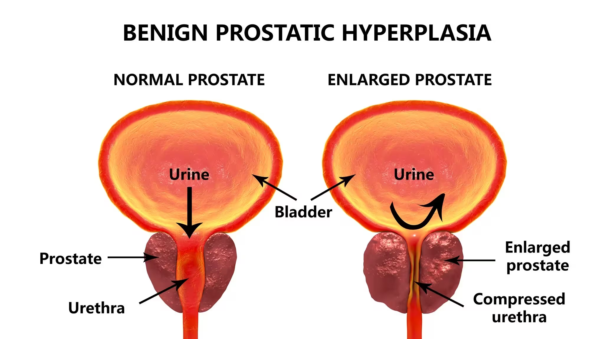

Benign prostatic hyperplasia, 3D illustration showing normal and enlarged prostate gland.
"Although BPH affects millions of men worldwide, scientists still do not fully understand why it develops primarily in the transition zone," senior author Vasan Yegnasubramanian said in the paper. "The molecular and zonal features of WIF1+ basal cells suggest they could contribute to processes that drive BPH."
When the authors cross-checked public datasets, the pattern persisted. Enlarged prostate samples showed enrichment of WIF1+ cells and a shifted gene signature compared with normal tissue. That strengthens the hypothesis that WIF1+ cells are not just bystanders but might actively shape the transition zone's susceptibility to hyperplasia.
What this could mean for diagnosis and treatment
The study stops short of proving causation. It offers a plausible cellular culprit and a molecular framework for future experiments. If WIF1+ basal cells are confirmed to drive regional prostate growth, they could become a new target for therapies designed to limit BPH without the systemic side effects of broad hormone suppression.
Targeting a defined cell population or the signaling pathways those cells use would be a more surgical approach at the molecular level. Functional studies will be needed, including cell lineage tracing and models that test whether manipulating WIF1 activity changes the trajectory of gland enlargement.
There is another clinical angle. Since the transition zone is relatively protected from cancer, understanding the mechanisms that both promote enlargement and suppress malignant transformation could help researchers separate pathways to benign growth from those that lead to tumors. That separation would be useful for screening and for developing safer treatments.
Expert Insight
"Finding a cell population that is anatomically and transcriptionally enriched in the transition zone is an important step," says Dr. Elena Martinez, a urologic researcher not involved with the study. "It gives us a testable target. The next phase should probe function: can we alter these cells in model systems to prevent or reverse enlargement? If so, the clinical implications could be substantial."

The Johns Hopkins results add texture to a medical problem that has been described clinically for generations but understood poorly at the cellular level. They do not rewrite the story overnight, but they give scientists a clearer map for where to look next: particular cells, localized interactions, and signaling pathways that together shape why an organ grows in one place and not another.

“My work centers on sustainability, energy, and environmental science — examining how innovation can lead to a greener future.”

Discussion
Leave a Comment
Comments
No comments yet. Be the first.