

A single brain offered researchers a rare, almost accidental experiment. Some regions were scrubbed nearly clean of amyloid plaques after years of anti-amyloid therapy. Others, only millimeters away, still carried heavy deposits. The contrast was stark. It exposed how removing amyloid in some places may arrest the cascade that leads to neuron loss while persistent amyloid elsewhere appears to allow other damaging processes to continue.
Uneven plaque removal created a natural experiment inside one patient’s brain. The findings suggest that where amyloid persists, other damaging processes may continue to advance.
Inside the case that changed a hypothesis into evidence
The brain belonged to a man in his fifties who had mild cognitive impairment from Alzheimer’s disease. He participated in a clinical trial and received 30 doses of aducanumab over four and a half years. He died roughly four years after his final dose and more than a decade after symptoms began. Before he passed, he donated his brain to the Center for Neurodegenerative Disease Research at Penn.
What the neuropathologists found was not uniform clearance. Brain imaging taken while the participant was alive had already hinted that plaque removal was patchy. The autopsy confirmed it and revealed the downstream effects. Raised ridges of brain tissue, called gyri, had lost most amyloid and showed only trace levels of abnormal tau protein. Adjacent grooves, the sulci, still harbored amyloid and had higher tau, more inflammation, and greater signs of neurodegeneration.
This spatial mismatch gave scientists a rare opportunity: to observe, within the same brain, how two different local environments evolved after anti-amyloid treatment. The uneven outcome is important because tau accumulation is more tightly linked than amyloid to where neurons die and where cognitive decline progresses. If removing amyloid can reduce tau spread in certain regions, that suggests a causal chain, not just coincidence.
What the researchers connected and why it matters
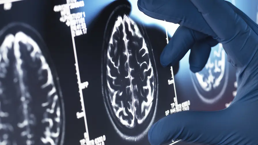
For years, the field has debated whether clearing amyloid is enough to slow Alzheimer’s. Clinical trials measure changes in cognition, brain scans, and fluid biomarkers. But those measures cannot reveal the detailed anatomy of protein spread the way an autopsy can. Autopsies from people treated with anti-amyloid drugs are exceptionally rare. That scarcity is why this single case matters.
The team from the Perelman School of Medicine at the University of Pennsylvania presented the results at the 2026 Alzheimer’s Association International Conference and published them in JAMA. They concluded that local amyloid clearance was associated with reduced tau pathology and less tissue damage in the same regions. The implication is straightforward. If plaques are removed in time and in the right places, the subsequent accumulation of tau and the inflammation that follows could be limited or delayed.
Edward B. Lee, one of the senior authors, described the case as a 'Goldilocks' scenario. Some areas cleared well. Others did not. That contrast allowed a direct comparison of what came after. David Wolk, another senior author and director at the Penn Memory Center, emphasized that observing both patterns in the same brain gave unusually clear human evidence that anti-amyloid therapies can influence downstream proteins that drive neurodegeneration.

Two brain regions show persistent amyloid plaque accumulation in the gyri, corresponding to greater tau tangle burden in the same areas. In contrast, the sulci exhibit greater amyloid clearance and, subsequently, reduced tau pathology. Top: Brain section from autopsy stained for beta-amyloid plaque. Bottom: Brain section from autopsy stained for tau tangles.
Timing, geography, and therapeutic strategy
Several critical questions arise from this case. How much plaque must be removed to translate into meaningful protection? Does the location of clearance matter more than the total amount removed? And crucially, how soon must treatment begin?

Clinical trials typically run for about 18 months. Disease biology does not always respect those timelines. The protective patterns seen in this brain unfolded over years, suggesting that some benefits of amyloid removal may appear slowly. If Alzheimer’s pathology develops silently for decades before symptoms surface, then a short trial may miss long-term advantages of amyloid targeting. That matters for trial design and for clinicians weighing when to treat.
The findings also raise a mechanistic question: why did the drug clear plaques more effectively from some brain regions than from others? Local differences in blood flow, immune activity, or the microenvironment of neurons could shape how antibodies reach and affect plaques. Understanding those pathways could inform modifications to existing drugs or the development of adjunct therapies that improve regional clearance.
These insights apply beyond one drug. Aducanumab was among the first anti-amyloid antibodies to reach patients, but newer agents such as donanemab and lecanemab have shown clearer clinical benefits in recent trials and now carry regulatory approvals. The Penn case suggests that the extent and pattern of amyloid reduction, not just whether any reduction occurs, may determine the therapy’s long-term protective effect against tau accumulation and neurodegeneration.
Expert Insight
'This case is a window into disease dynamics we can rarely observe,' said Dr. Maya Lin, a fictional neurologist and neuroimmunology researcher at a major academic center. 'It tells us that Alzheimer’s is not uniform across the brain. Therapies will likely need to be evaluated by where and when they act, not just by global measures. The next step is mapping why some regions respond and others do not, and then designing treatments that reach vulnerable pockets early enough to prevent tau from taking hold.'
Dr. Lin’s perspective underscores a practical point for ongoing and future trials: longer follow up, higher-resolution imaging, and post-mortem donations will be essential to translate molecular changes into clinical decisions. Building tissue donation into trial protocols could accelerate the science.
Conclusion

This single autopsy does not prove a universal rule. It does not answer how much plaque removal is enough or guarantee that every person treated will see this pattern. Yet it provides one of the clearest human demonstrations to date that removing amyloid can influence the biological cascade that leads to tau buildup and neural damage. The case argues for earlier detection, for careful mapping of regional responses, and for patience when assessing the long-term benefits of anti-amyloid therapies. Continued studies, including longer clinical follow up and additional brain donations, will be essential to move from hopeful signal to reliable strategy.

“The cosmos has always fascinated me. I write about space missions, astronomy, and the technologies pushing humanity beyond Earth.”

Discussion
Leave a Comment
Comments (3)
Interesting, but feels a bit overhyped. one brain is neat evidence, yet not definitive. also why did some regions resist clearance? 🤔 gotta map that more.
wait, one case only tho? is this enough to change practice? sounds promising but still need more autopsies, longer follow up...
wow, that inside-brain contrast is wild. patchy plaque removal? hopeful but kinda scary. timing looks everything, trials too short imho