
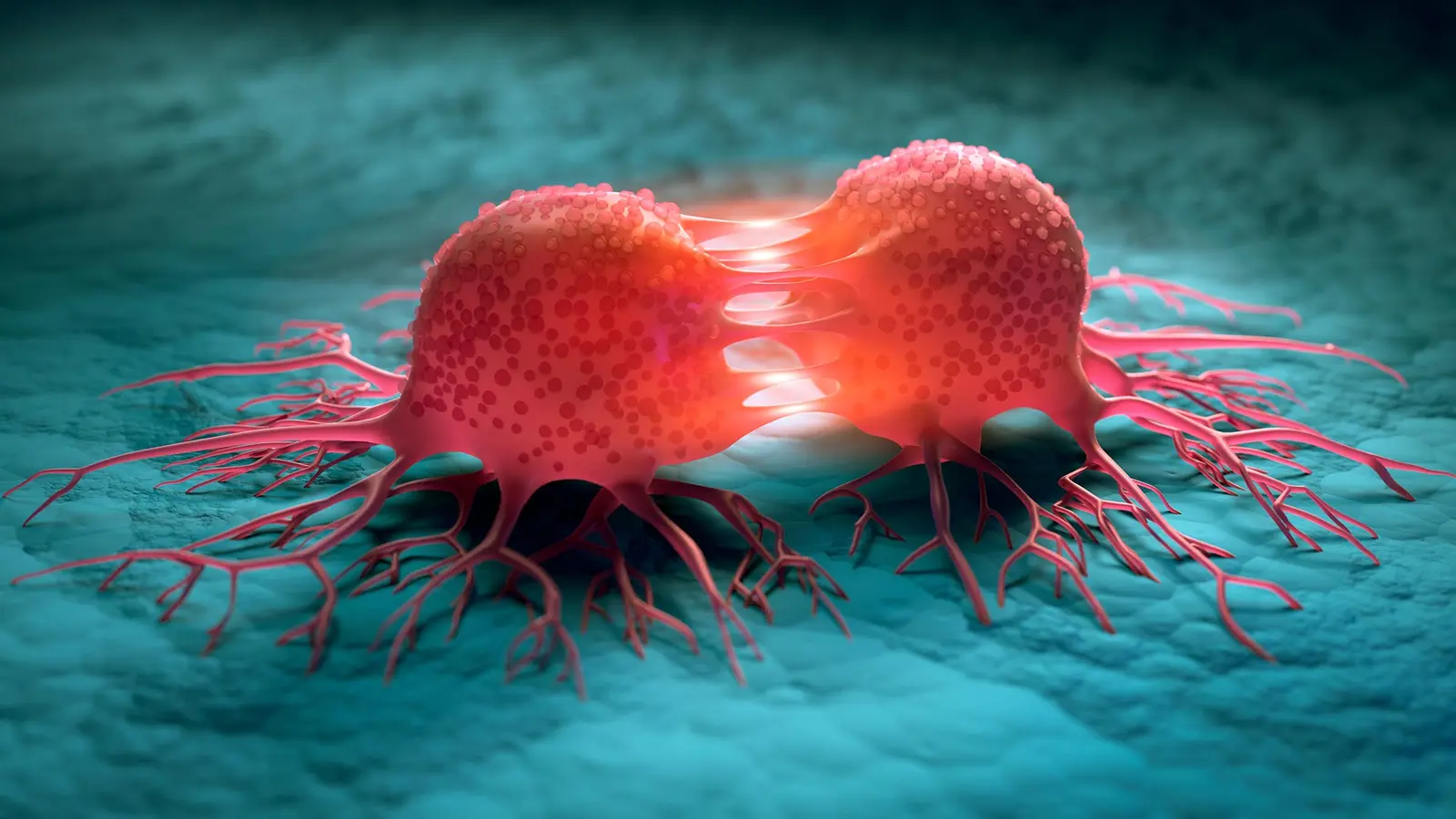
Imagine a microscopic crossroads where human bone, lung and blood vessels meet, and where cancer makes the risky leap from circulation into a new home. That is precisely what researchers at Columbia University have built: a multi-organ, human-derived chip that lets scientists watch circulating breast cancer cells exit the bloodstream and begin the earliest, most secretive phase of metastasis.
Why this matters now
Cancer becomes lethal not when the primary tumor grows, but when cells depart that origin and seed distant organs. Most cancer deaths trace back to metastasis, yet therapies designed specifically to stop or slow this cascade have proven stubbornly elusive. One reason is simple: we lack accurate, predictive human systems that reveal how metastatic cells behave in a human environment. Rodent models taught us a great deal, but mouse physiology is not a perfect stand-in for the human body. That mismatch helps explain why some drugs that look promising in animals fail in clinical trials.
The Columbia chip addresses a narrow but crucial slice of the metastatic journey. It recreates organ colonization, the stage when tumor cells attach to the inner lining of blood vessels, cross that barrier, survive hostile tissue conditions and eventually expand into secondary tumors. Observing those events in real time, inside human tissues, opens a new window on how cancers choose and prepare their targets.
How the multi-organ chip is built
The system uses three engineered human tissues: bone, lung and vascular endothelium. All originated from induced pluripotent stem cells, or iPSCs, which researchers coax into specific cell types and then nurture inside tissue-specific scaffolds and bioreactors so they mature and function like the real thing. The tissues occupy separate millimeter-scale compartments linked by a flowing vascular channel.
Design choices that matter
- Separate but connected compartments: each organ retains its own microenvironment while remaining exposed to a shared circulatory flow.
- A selectively permeable endothelial barrier: this mimics blood vessel walls and lets researchers track how tumor cells adhere, traverse and invade.
- Patient-derived cell compatibility: the chip can accept cells from different people, enabling personalized studies of metastatic behavior.
The platform does more than place tissues beside tumor cells. It recreates the continuous biochemical dialogue between circulating cancer cells and target organs. That conversation determines whether a stray cell is repelled, destroyed, or allowed to take root.

The Columbia team modeled how cancer spreads to other organs using lab-grown bone and lung tissues.
What the experiments revealed
When researchers introduced circulating breast cancer cells into the vascular channel, the device reproduced known patterns of organ preference, or organ tropism. Breast cancer cells that in patients tend to colonize bone did so more aggressively in the engineered bone compartment, leaving greater tissue damage. Cells with a predilection for the lung caused more harm in the lung compartment while only weakly invading bone. Those differences showed that the chip captures organ-selective invasion and the secretion profiles that accompany tissue-specific colonization.
Another striking result: tumor cells altered distant tissues before mass colonization occurred. This pre-metastatic conditioning means a cancer can send molecular signals ahead of migrating cells, remodeling a future landing zone to favor survival and growth. The chip reproduced aspects of that incubation process, giving researchers a platform to study how and when these early changes occur and—crucially—how they might be interrupted.
Lead investigator Gordana Vunjak-Novakovic described the motivation behind the work as a gap in human-relevant tools for studying metastasis. Paraphrasing her remarks, the goal was to observe whether cancer cells can adhere to and cross endothelium and then to test their ability to survive and reprogram the tissue they invade. The platform allowed the team to do precisely that, tracking physical crossing events and the downstream remodeling of the local niche.
Expert Insight
Dr. Elena Márquez, a translational oncology researcher not involved in the study, offered this real-world perspective: 'What makes this chip important is its human cellular context combined with flow dynamics. Blood flow and endothelial interactions shape metastatic fate in ways static cultures cannot reproduce. A device like this helps bridge laboratory discovery and patient-relevant biology.' Her comment highlights the potential for this approach to inform which preclinical findings are worth translating toward human trials.
Implications for drug discovery and personalized medicine
Organs-on-chips are not a wholesale replacement for animal studies. Instead, they can fill critical blind spots, especially when researchers need human-specific responses. By permitting controlled perturbations—alter one tissue component, swap in patient-derived tumor cells, or inhibit candidate molecular pathways—investigators can prioritize targets that are more likely to work in people.
The Columbia work also lines up with regulatory and research trends. Agencies such as the FDA and NIH are increasingly encouraging 'new approach methodologies' that reduce reliance on animal testing and improve human predictive power. A human, patient-adaptable platform that reproduces organ tropism and pre-metastatic niche formation provides a concrete example of how such methodologies might function in practice.
Collaborations across disciplines strengthened the project. The effort brought together tissue engineers, cancer systems biologists and pathologists to combine microphysiological engineering with molecular profiling and clinical insight. That multidisciplinary approach matters because metastasis is not a single-cell problem. It is a complex, adaptive interaction among tumor cells, vasculature and resident tissue cells.
Conclusion
The Columbia multi-organ chip marks a meaningful step toward observing the earliest stages of organ colonization in human tissue. It gives researchers a controlled, humanized environment to study organ-specific invasion, pre-metastatic conditioning and the molecular circuits that give metastatic cells an edge. These capabilities could help prioritize new therapeutic targets, refine preclinical testing, and ultimately move more effective interventions into clinical trials faster.
There remains work to do. Chips cannot yet capture the full complexity of whole-body physiology: immune system diversity, long-range endocrine signals and the many cell types that define each organ. Still, by shining a light on a formerly opaque phase of cancer spread, this platform provides a sharper lens on one of oncology's toughest challenges. The next step is to use that lens to test interventions that might stop metastasis before it starts.

“The cosmos has always fascinated me. I write about space missions, astronomy, and the technologies pushing humanity beyond Earth.”

Discussion
Leave a Comment
Comments (3)
I worked with iPSC endothelium, this rings true, but scaling up and immune parts? that's the hard part, hope they tackle it soon.
Is this even true? Looks cool but can a chip really mimic immune diversity and long-term signals? skeptical but hopeful.
Wow, seeing metastasis in human tissue chips gives me chills. If it pans out it could change trials, but need more validation..