
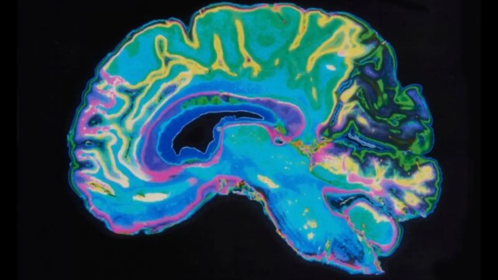
Picture a brain that, for a time, seems to stop shrinking. Not a reversal. Not rejuvenation. Just a pause. That is the striking observation at the center of a new longitudinal MRI study that tracked more than 1,000 people through life’s major biological transitions.
A surprising stall in cortical shrinkage
Neuroscientists led by Sophie van 't Hof at Amsterdam University Medical Center reported what no one had measured before: during the transition into menopause the steady, age-related decline in cortical gray matter volume slows. The work, published in Nature Communications in 2026, compared brain scans taken repeatedly across cohorts experiencing puberty, pregnancy, and menopause. Each phase showed distinct patterns. Puberty and pregnancy correlated with accelerated cortical thinning. Menopause did not.
The finding is counterintuitive because cortical volume typically falls with age. Yet when participants entered the menopausal transition, that decline appeared to stall. The effect was not tied to chronological age alone. It showed up specifically in the group whose reproductive status changed during the study window; participants who were pre-menopausal or post-menopausal for the entire study continued the expected pattern of gray matter loss.
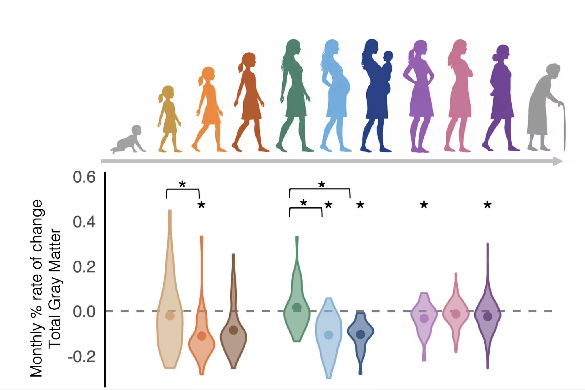
The female brain shows changes to total gray matter across a lifetime.
Study design in brief
- More than 1,000 individuals underwent repeated MRI scans over several years.
- Researchers sampled people at life stages with large hormonal shifts: puberty, pregnancy, and the menopause transition.
- Participants self-reported menopausal stage; blood hormone measures were not collected.
- Analyses accounted for variables such as age, hormone replacement therapy use, and number of children.
Repeated imaging strengthens the claim. Scans show temporal associations between life stage and brain-volume change. They do not prove causation. Still, the pattern stands: the menopause transition, unlike puberty or pregnancy, coincides with an attenuation of whole-brain cortical gray matter loss.
Where in the brain did these changes cluster? Regions known to be sensitive to estrogen signaling showed notable effects, although estrogen receptors are widespread. That concordance hints at a hormonal component. But because hormone concentrations were not measured, the link remains speculative. Correlation, not causation.
Previous work complicates the picture. A 2018 longitudinal study that focused on the hippocampus reported accelerated hippocampal volume loss across the menopause transition. The new, broader cortical analysis paints a different portrait: possible slowing of age-related decline in total cortical volume during the transition. The authors caution this interpretation is preliminary, and rightly so.
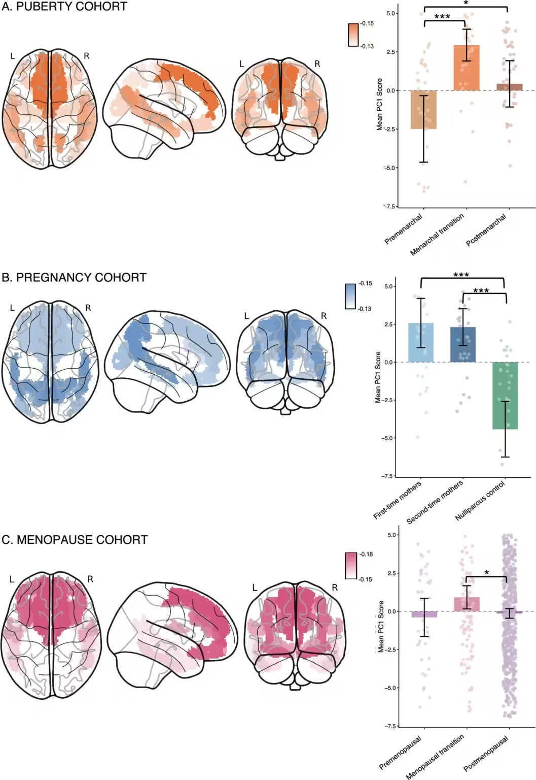
Top 20 regions with the highest loading of the principal component analysis per cohort and distribution of PC1 scores across groups for the A: puberty, B: pregnancy, and C: menopause cohorts.
What this could mean for brain aging
Biology does not always move in straight lines. Cortical thinning during adolescence and pregnancy is thought to reflect adaptive reorganization: pruning and reshaping to match new cognitive and bodily demands. Could menopause trigger another kind of reorganization, a temporary redistribution of neural resources as sex-steroid levels change?
We do not have a firm answer. The study shows an association that survives tests for confounding factors such as age, hormone therapy use, and parity. It also highlights a gap in neuroscience: female-specific longitudinal research is rare. Only a small fraction of studies focus exclusively on females or treat sex as a core analytic variable. That scarcity slows our ability to build clear mechanistic narratives about how hormonal transitions sculpt the aging brain.
Expert Insight
'This work opens an intriguing window on brain plasticity during midlife,' says Dr Elena Morales, a clinical neurologist who studies hormonal influences on cognition. 'If cortical loss slows during menopause, we need to know whether that pause is protective, neutral, or a prelude to later changes. Longitudinal hormone sampling and functional measures will be critical next steps.'
Dr Morales points out practical priorities. 'Combine MRI with blood or saliva hormone profiles. Track cognitive trajectories alongside structural change. Include diverse populations. Only then can we separate biological signal from noise.'
Conclusion
The new study does not settle whether hormonal declines cause the observed cortical slowdown, or whether that slowdown has positive or negative cognitive consequences. What it does do is spotlight an unexpected pattern: menopause appears to be defined less by accelerated loss and more by a distinct alteration in the brain’s aging trajectory. For clinicians and researchers, the message is clear. Female-specific, longitudinal neuroscience deserves more attention and richer measurements.

Menopause appears associated with a temporary slowing of cortical gray matter loss.

“My work centers on sustainability, energy, and environmental science — examining how innovation can lead to a greener future.”

Discussion
Leave a Comment
Comments (1)
Wait so menopause slows cortical loss? hmm, without hormone measures that sounds premature. Need blood tests + cognitive followup, not just MRI.